Epitope of rheumatoid arthritis and application thereof

An antigen epitope and arthritis technology, applied in the field of immunological diagnosis, can solve the problems of unclear pathogenesis, self-antigen detection antibody, etc.

Inactive Publication Date: 2012-11-28
INST OF BASIC MEDICAL SCI ACAD OF MILITARY MEDICAL SCI OF PLA +1
View PDF3 Cites 22 Cited by
  • Summary
  • Abstract
  • Description
  • Claims
  • Application Information

AI Technical Summary

Problems solved by technology

[0004] The appearance of a large number of autoantibodies is one of the important characteristics of autoimmune diseases, but because its pathogenesis is still unclear, it is difficult to directly use autoantigens to detect antibodies

Method used


Image

  • Epitope of rheumatoid arthritis and application thereof
  • Epitope of rheumatoid arthritis and application thereof
  • Epitope of rheumatoid arthritis and application thereof

Examples


Embodiment 1

[0020] Example 1 Preparation and purification of RA patient serum total IgG

[0021] Take an appropriate amount of Protein A resin suspension (product of GE Company), put it into the chromatography column, and wash the equilibrium chromatography column with 10 times the column volume of PBS buffer. After centrifuging the serum of patients with rheumatoid arthritis at high speed, add 1 / 10 volume of 10×PBS buffer solution to mix, adjust the pH and ion concentration, and slowly add to the chromatography column. Wash with more than 10 times the column volume of PBS until no protein is detected in the effluent. Add 6 times column volume of 0.1 M glycine (pH 3.0) to elute, collect the flow-through, and neutralize with 1 / 10 volume of 1M Tris-HCl (pH 9.0). Dialyze the obtained antibody against PBS, add 50% glycerol after quantifying the antibody concentration, aliquot and store at -20°C.

Embodiment 2

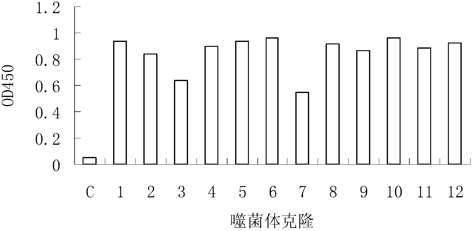
[0022] Example 2 Phage display peptide library screening for RA-specific antibody-binding peptides

[0023] Ph.D.-12 peptide library kit was purchased from NEB Company, with a library capacity of 2.7×10 9 , with a titer of 1.5×10 13 / μl.. Escherichia coli ER2537 is the host strain of the peptide library. For the screening process, refer to the instruction manual of the phage display peptide library kit. Each round of screening was coated with total IgG in RA patient serum (10 μg per well), and 1 × 10 11 Phage. The degree of phage enrichment was calculated by "input / output ratio". After three rounds of screening, positive clones were selected for sequencing. The specific binding of positive phage clones to total IgG antibodies was determined by ELISA. Total IgG antibody (10 μg per well) was coated on a 96-well plate (Nunc Company) overnight at 4 °C. Pour off the unbound antibody and block with 3% BSA at 37°C for 1 hour. Put 1×10 in each hole 9 Phage were incubated at ...

Embodiment 3

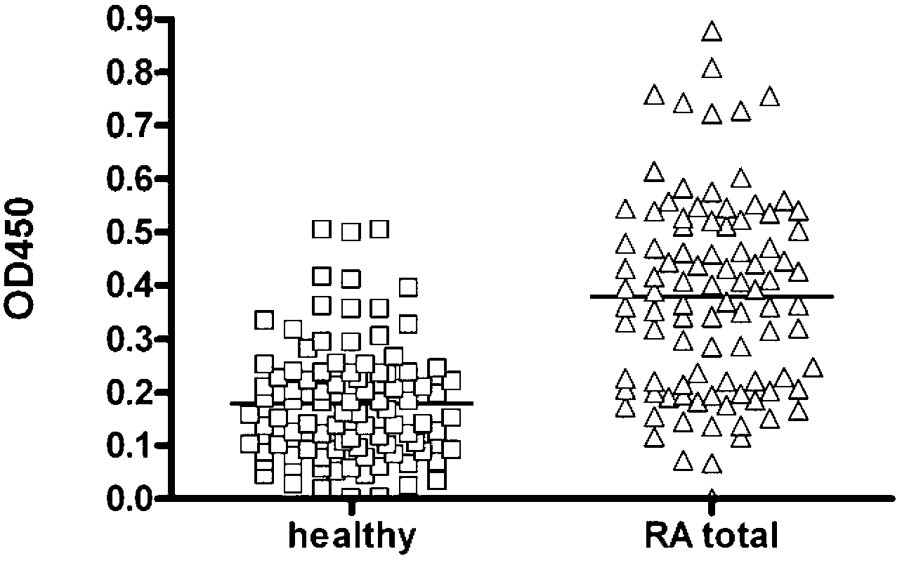
[0024] Example 3 Detection of the combination of RAP and RA patient serum IgG

[0025] The synthetic peptide RAP (0.5 μg per well) was coated on a 96-well plate (Nunc Company) overnight at 4 °C. Unbound peptides were poured off and blocked with 3% BSA at 37°C for 1 hour. Serum samples from RA patients or healthy individuals were diluted 1:400, added to different wells, and incubated at 37°C for 1 hour. Wash the plate with washing solution (PBS-0.05% Tween 20) 5 times for a total of 3 minutes. 1:1000 dilution of HRP-labeled goat anti-human IgG was incubated at 37°C for 1 hour. The washing method was the same as above, and the bound anti-M13 monoclonal antibody was detected with tetramethylbenzidine (TMB, Sigma) as the substrate, and the light absorption was detected at 450 nm.

[0026] see results figure 2 , the level of RAP antibody in the serum of RA patients was significantly higher than that of healthy people. Statistical analysis was performed using One-way ANOVA meth...


PUM


Abstract

The invention discloses an epitope of rheumatoid arthritis and application thereof, belonging to the technical field of immunologic diagnosis. The epitope is a polypeptide, and the amino acid sequence is disclosed as SEQ ID NO:1 in the sequence table. ELISA detection and patient serum reaction condition indicate that the epitope polypeptide can be specifically combined with IgG in the patient serum, and does not react with the serum of a health person. The epitope polypeptide can be used for preparing medicines for diagnosing rheumatoid arthritis.

Description

technical field [0001] The invention belongs to the technical field of immunological diagnosis, and in particular relates to an antigenic epitope of rheumatoid arthritis and its application. Background technique [0002] Rheumatoid arthritis (RA) is a systemic autoimmune disease based on joint synovial inflammation and bone destruction. The prevalence rate in my country is 0.34%. There are more than 5 million RA patients in China, and Most of them are young and middle-aged laborers, and it is a major disabling disease that seriously endangers human health. So far, the pathogenesis of this disease is still unclear, and the treatment is mainly non-specific symptomatic treatment, which temporarily relieves and improves the condition, but cannot fundamentally control the occurrence of synovial inflammation and the progress of bone destruction. [0003] In recent years, a citrulline-related autoantibody system with high specificity for the diagnosis of RA has been proposed, inclu...

Claims


Application Information

Patent Timeline
Patent Type & Authority Applications(China)
IPC IPC(8): C07K7/08C12N15/12G01N33/68
Inventor 杨光栗占国高亚萍柳川穆荣董洁刘玉
Owner INST OF BASIC MEDICAL SCI ACAD OF MILITARY MEDICAL SCI OF PLA
Why Eureka
Try Eureka