A nano-microsphere time-resolved fluorescent probe and its preparation method and application
A time-resolved fluorescence, nano-microsphere technology, applied in the field of detection, can solve the problem of insufficient fluorescence intensity of rare earth element fluorescent probes, and achieve the effects of large compatibility of detection samples, rapid and quantitative combination, and high fluorescence intensity
- Summary
- Abstract
- Description
- Claims
- Application Information
AI Technical Summary
Problems solved by technology
Method used
Image

Examples
Embodiment 1
[0021] Embodiment 1 Preparation of carboxylated polystyrene nanospheres
[0022] Dissolve 10mm of styrene monomer and 0.95mm of acrylic acid monomer in 10ml of deionized water containing 0.45mm of sodium dodecylsulfonate, add it to a round bottom flask, stir evenly with a magnetic stirring bar, and then use high-purity nitrogen gas to Remove all the air in the bottom flask, seal and heat to 70°C, add 0.5ml of 0.15mm potassium persulfate, seal and stir for 8 hours in an oxygen barrier, then cool down to room temperature, then filter the reaction solution with whatman 2v filter paper (pore size 8 μm), Finally, a dialysis bag (molecular weight cut-off: 30,000 Da) was used for dialysis against deionized water for 5 days, and the liquid in the dialysis bag was collected and stored at 4° C. by adding 0.05% sodium azide.
[0023] By measurement, the prepared carboxylated polystyrene nanospheres have a diameter of 190 ± 10nm, a surface charge Surface Charge (μeq / g) of 170-200, and a c...
Embodiment 2
[0024] The preparation of embodiment 2 nano fluorescent microspheres
[0025] Get a small amount of 190nm polystyrene microspheres prepared in Example 1, add in 10ml of deionized water and acetone mixed solution (v / v=1:1), make the density of polystyrene microspheres in the reaction solution be about 1 ×10 14 Each, stir well, add 100ul 0.1M europium trichloride, 1 μl 0.1M terbium trichloride, 400 μl 0.1M β-diketone (β-NTA), 300 μl trioctylphosphine oxide (TOPO), 100 μl phenanthroline, First heat to 60°C and stir at constant temperature to avoid light for 10 hours, then lower to room temperature and react for 2 hours, finally remove the organic solvent in the solution by vacuum distillation, dialyze against deionized water for 5 days, remove the remaining small molecular substances, collect and dialyze The liquid in the bag was stored at 4°C by adding 0.05% sodium azide.
[0026] Through testing and calculation, the average number of europium ion chelates wrapped in each nano...
Embodiment 3
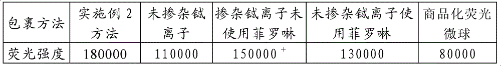
[0027] Embodiment 3 Comparison of fluorescence intensity of nano-fluorescent microspheres by different wrapping methods
[0028] Refer to the formula and steps of Example 2 to prepare undoped terbium ions, doped terbium ions without using phenanthroline, and undoped terbium ions using phenanthroline nano-fluorescent microspheres, and compared the fluorescence intensity. The results are shown in Table 1 .
[0029] Table 1
[0030]
[0031] Note: (1) Fluorescence intensity is defined as the multiple of the fluorescence intensity produced by a nanosphere after excitation that is equivalent to that of a single free europium ion chelate;
[0032] (2) Commercial fluorescent microspheres with a particle size of 0.2 μm were purchased from Thermo Fisher Scientific under the trade name of Fluoro-Max Carboxylate-Modified and Streptavidin-Coated Europium Chelate Particles.
PUM
| Property | Measurement | Unit |
|---|---|---|
| diameter | aaaaa | aaaaa |
| particle diameter | aaaaa | aaaaa |
Abstract
Description
Claims
Application Information