High-sensitivity protein detection method

A protein detection and high-sensitivity technology, applied in the field of high-sensitivity protein detection, can solve problems such as difficult and fast washing, affecting sensitivity, and increasing the background of magnetic beads

Inactive Publication Date: 2015-03-18
HANGZHOU JINXI BIOLOGICAL TECH
View PDF4 Cites 2 Cited by
  • Summary
  • Abstract
  • Description
  • Claims
  • Application Information

AI Technical Summary

Problems solved by technology

However, there are also some problems in the magnetic bead solid-phase PLA that affect its use.
First of all, due to the presence of magnetic beads, when performing fluorescent quantitative PCR, the magnetic beads will increase the background and affect the sensitivity of the detection; secondly, during the washing process, not all the magnetic beads will be captured by the magnetic frame. Washing the magnetic beads will reduce some, which will affect the sensitivity of the detection, resulting in large error and poor stability; in addition, the washing technology of magnetic beads requires high requirements, such as magnetic frame and other equipment, it is difficult to wash quickly, the process is slow, and it is difficult to achieve high-throughput of samples. Quantity detection
These problems have affected the promotion and use of solid-phase PLA
[0008] Therefore, although PLA has high sensitivity as a new protein detection technology, there are still many problems affecting its use, and it is difficult to use this method to produce products for the detection of diseases and other fields to serve the society

Method used


Image

  • High-sensitivity protein detection method
  • High-sensitivity protein detection method
  • High-sensitivity protein detection method

Examples


Embodiment 1

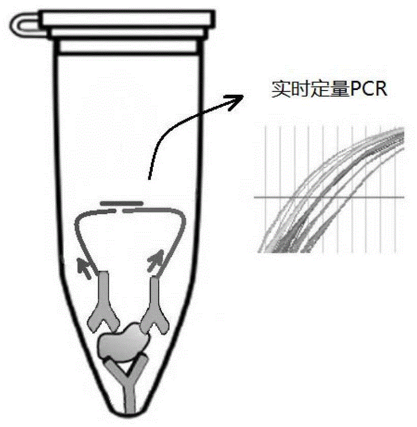
[0078] Embodiment 1. A high-sensitivity protein detection method, using the direct coating method of mode 1 to carry out solid-phase PLA on the tube wall, specifically the following steps:

[0079] 1) Add 50 ul of PAB240 antibody at a concentration of 0.1 ug / ml to each quantitative PCR tube, place it at 4°C for 12 hours, and wash with PBST buffer 3 times (each volume is 200 ul); the purpose of washing It is to wash away the antibody that is not bound to the tube wall.

[0080] 2) Add 200 ul of BSA solution with a concentration of 10g / L to each of the quantitative PCR tubes coated with antibodies above (that is, each quantitative PCR tube obtained in step 1), and discard the BSA solution after standing at 37°C for 2 hours , after drying at 25 ℃, put it at 4 ℃ for later use; the shelf life at 4 ℃ can reach 6-12 months.

[0081] 3) 100 microliters of two biotinylated single-stranded DNAs (probe DNA1 and probe DNA2) with a concentration of 100 nanomoles / liter, and carry out the f...

Embodiment 2

[0112] Embodiment 2. A high-sensitivity protein detection method, using the cross-linking agent immobilization method of mode 2 to carry out solid-phase PLA on the tube wall, specifically:

[0113] Insert the following prologue steps before step 1) of Example 1:

[0114] Add 50 ul / tube of cross-linking agent solution (glutaraldehyde solution with volume concentration of 1%) to the quantitative PCR tube, place it at 37°C for 5 hours, and wash it with deionized water for 3 times (200 ul for each time; The purpose is to remove the unreacted cross-linking agent in the PCR tube); get the quantitative PCR tube after pretreatment.

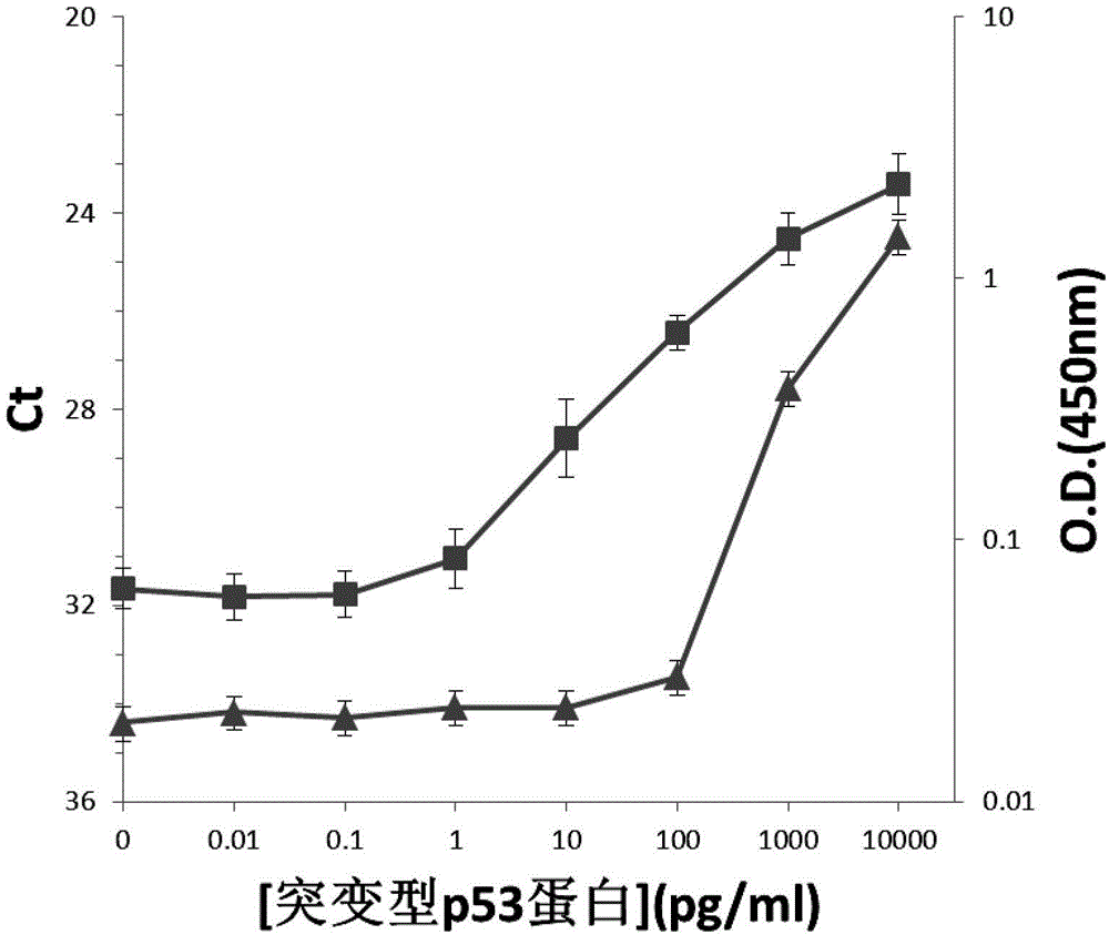
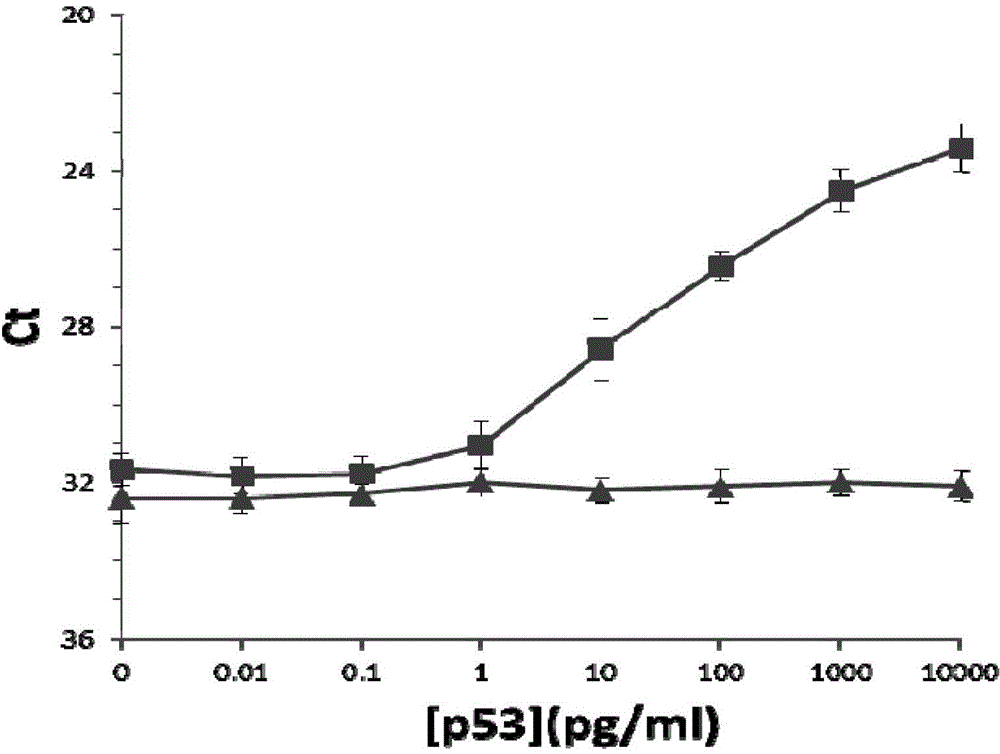
[0115]Then use this pre-treated quantitative PCR tube to replace the quantitative PCR tube in Step 1) of Example 1, and the rest are the same as in Example 1. At the same time, the pre-treated quantitative PCR tube was used instead of the quantitative PCR tube in Step 1) of Example 1, as in verification test 2 in Example 1, to detect the normal P53 prote...

Embodiment 3

[0125] Example 3. Detection of cardiac troponin cTnI

[0126] Cardiac troponin cTnI in human serum is closely related to myocardial infarction. We use cross-linking agent immobilization method of mode 2 with higher detection sensitivity to detect cardiac troponin cTnI by solid-phase PLA on the tube wall. That is, according to Example 2, 50 ul / tube of a cross-linking agent solution (glutaraldehyde solution with a volume concentration of 1%) was added to the quantitative PCR tube, and after standing at 37°C for 5 hours, it was washed 3 times with deionized water (each The amount used is 200 ul; the purpose of washing is to remove the unreacted cross-linking agent in the PCR tube); the quantitative PCR tube after pretreatment is obtained. Then use this pre-treated quantitative PCR tube to replace the quantitative PCR tube in step 1) of Example 1, but use cTnI monoclonal antibody (purchased from American R&D Company, catalog number: ab47003) to replace the p53 mutant monoclonal a...


PUM

PropertyMeasurementUnit
Sensitivityaaaaaaaaaa

Abstract

The invention discloses a high-sensitivity protein detection method, wherein one of the following modes is selected optionally: mode 1, performing a tube wall solid phase PLA (proximity ligation assay) by a direct coating method, namely, directly adsorbing the protein or the antibody to a real-time fluorescence quantification PCR (Polymerase Chain Reaction) tube wall to perform PLA detection; and mode 2, performing a tube wall solid phase PLA by a cross-linking agent fixing method, namely, cross-linking the protein or the antibody to the real-time fluorescence quantification PCR tube wall by glutaraldehyde to perform PLA detection. Specifically, the method comprises the following steps of: using a monoclonal antibody pab240, and sequentially establishing PLA detection of P53 protein (P53 protein mutant) and cTnI (cardiac troponin I). According to the method disclosed by the invention, the antibody is fixed on a PCR tube so as to perform protein PLA detection, magnetic beads are not needed in the detection process, defects caused by application of the magnetic beads are overcome, and the detection sensitivity to the protein is also improved greatly.

Description

technical field [0001] The invention relates to a highly sensitive protein detection method. Background technique [0002] Proximity ligation assay (PLA) technology is a protein detection technology developed by Fredriksson et al. after 2000. Originally, the probes used in the PLA technique could also be single strands of DNA called aptamers. The nucleic acid aptamer is screened by an in vitro screening method, and can combine with a target molecule with high specificity and high affinity, so as to recognize the target molecule. Nucleic acid aptamers are easy to synthesize and easy to store, and have been widely used in basic research, diagnosis, development of therapeutic reagents, drug screening and other fields. However, the types of nucleic acid aptamers that can be used in PLA technology are currently limited, which greatly limits the application of nucleic acid aptamers in this technology. [0003] At present, PLA technology has been greatly developed. To detect pr...

Claims


Application Information

Patent Timeline
Patent Type & Authority Patents(China)
IPC IPC(8): C12Q1/68G01N33/68G01N33/577
Inventor 朱成钢王英杰易文江学成
Owner HANGZHOU JINXI BIOLOGICAL TECH
Why Eureka
Try Eureka