Quantitative detection device based on fibrous-membrane gathering and separation and detection method thereof

A technology of quantitative detection and fiber membrane, applied in the direction of measuring devices, analysis materials, instruments, etc., can solve the problem of low accuracy of quantitative detection

Active Publication Date: 2013-09-25
SHANGHAI YUNZE BIOTECH +1
View PDF17 Cites 30 Cited by
  • Summary
  • Abstract
  • Description
  • Claims
  • Application Information

AI Technical Summary

Problems solved by technology

[0006] The invention provides a special device for quantitative and detection methods based on fiber membrane capture and separation, which is mainly used to solve the technical problem of low quantitative detection accuracy of existing IF and IC technologies; the met

Method used


Image

  • Quantitative detection device based on fibrous-membrane gathering and separation and detection method thereof
  • Quantitative detection device based on fibrous-membrane gathering and separation and detection method thereof
  • Quantitative detection device based on fibrous-membrane gathering and separation and detection method thereof

Examples


Embodiment 1

[0082] Embodiment 1 uses BHTCT-Eu 3+ Implement IP-10TRIFMA for markers

[0083] After kidney transplantation, with the improvement of the transplanted kidney function, the urine IP-10 concentration tends to decrease significantly; the determination of urine IP-10 is a diagnostic index for monitoring the transplanted kidney function.

[0084] Proceed as follows:

[0085] 1) Preparation of capture antibody

[0086] A) 0.3 mg of anti-IP-10 monoclonal antibody (Lifespan Biosciences, Inc.) was dialyzed overnight against 500 ml of PBS buffer (50 mM, pH 8.0, containing 0.9% NaCl).

[0087] B) Add 1ml MES buffer (50mM, pH6.5) to 200μl latex microspheres (ACME microspheres, USA, Catalog No.CP255-10), mix well, centrifuge at 5000×g for 20min, remove the supernatant, and add 1.2ml Disperse in MES buffer; add 200 μl of MES buffer containing 4 mg EDCA and 8 mg Sulfo-NHS, and react at room temperature for 20 min.

[0088] C) Centrifuge at 5000×g for 20 min, discard the supernatant; add ...

Embodiment 2

[0112] Embodiment 2 HBV DNA hybridization analysis

[0113] HBV infection after liver transplantation is an important topic in liver transplantation surgery in my country. Routine serological testing is often unable to determine the HBV infection status of liver donors, and some HBsAg-negative donors still contain low levels of HBV replication and are infectious to a certain extent. Therefore, the determination of HBV DNA in organ donors can supplement the lack of serological testing , to avoid the occurrence of HBV reinfection after liver transplantation.

[0114] 1) Sample DNA extraction

[0115] The HBV DNA template was extracted using the QIAamp MinElute Virus Spin kit (QIAGEN, Germany): add 200 μl buffer AL and 25 μl QIAGEN protein to a test tube containing 200 μl serum, incubate at 56°C for 15 min, add 250 μl absolute ethanol, and incubate at room temperature for 5 min. Add the enzymatic solution to the separation microcolumn, centrifuge at 6000g for 1min; add 500μl bu...


PUM


Abstract

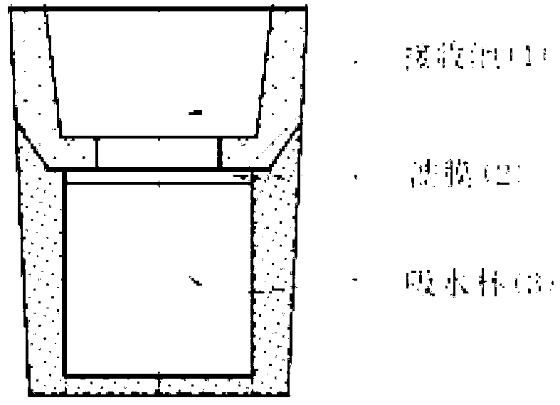
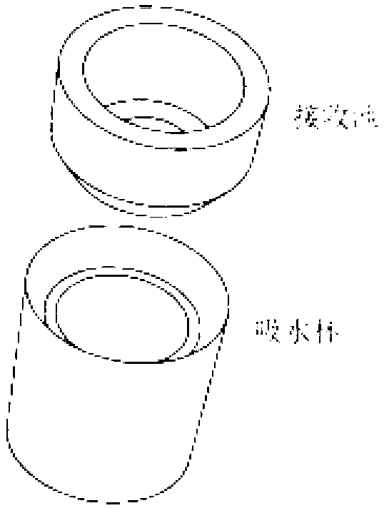
The invention relates to a quantitative detection device based on fibrous-membrane gathering and separation and a detection method thereof. The quantitative detection device comprises a separation testing cup, a reaction cup, an analysis buffer solution and a cleaning solution; wherein the separation testing cup comprises a receiving tank, a water absorption cup and millipore filters between them. A capture agent and a labeling agent are solidified on the bottom of the reaction cup and the capture agent is fixed on the surface of latex beads. The detection method comprises: conjugation reaction, separation, cleaning and detection. In the invention, not only the characteristics of convenient operation and rapid detection of immune infiltration (IF) and immunochromatography (IC) are maintained, but also precision and sensitivity of the detection are obviously better than that of IF and IC. Therefore, quantitative immunodetection and nucleic acid hybridization analysis can be implemented effectively. The invention can be used for the quantitative detection of substances such as protein, nucleic acid, small molecule and microbe.

Description

technical field [0001] The invention belongs to the field of biological analysis devices and detection methods thereof, in particular to a quantitative detection device and detection method based on fiber membrane capture and separation. Background technique [0002] Since the 1960s, bioanalysis technologies represented by immunoassay and nucleic acid analysis have been widely used in various fields such as clinical diagnosis, drug monitoring, food / environmental monitoring, and epidemiological investigation, and have become indispensable in people's production, life and scientific practice. Missing detection tools. With the advancement of antibody / antigen preparation technology, the development of various high-sensitivity detection technologies, nucleic acid amplification technology, and the improvement of the automation level of detection equipment, in recent years, the development of bioanalysis has gradually shifted to central laboratory detection and POCT detection (Poin...

Claims


Application Information

Patent Timeline
IPC IPC(8): G01N33/558G01N33/543
Inventor 史小娟孙宏彬乜永艳
Owner SHANGHAI YUNZE BIOTECH
Why Eureka
Try Eureka