Specific a1at monoclonal antibody for detection of endometriosis
A monoclonal antibody and detection kit technology, applied in the field of detection and/or detection of endometriosis, can solve the problems of delaying the timing of doctor's inspection, insufficient sensitivity of endometriosis, and low patient acceptance
- Summary
- Abstract
- Description
- Claims
- Application Information
AI Technical Summary
Problems solved by technology
Method used
Image

Examples
Embodiment 1
[0058] Example 1 Preparation of Antigen
[0059] The samples collected in this embodiment were women with endometriosis, and their serum samples were collected after informed consent. Serum samples were precipitated with 4 volumes of ice-cold acetone (containing 10% (w / v) trichloroacetic acid (TCA)). The mixture was placed at -20°C for 90 minutes, then centrifuged at 15,000 xg for 20 minutes at 4°C to collect the precipitate. The precipitate was then washed with ice-cold acetone, and centrifuged again at a speed of 15,000×g for 20 minutes. Remove the supernatant and wash with rehydration buffer (7M urea, 4% CHAPS ([3-(3-cholaminopropyl) dimethylamino]-1-propanesulfonate), 2M Thiourea (thiourea), 0.002% bromophenol blue (bromophenol blue) and 65mM dithioerythritol (DTE) dissolved the precipitate.
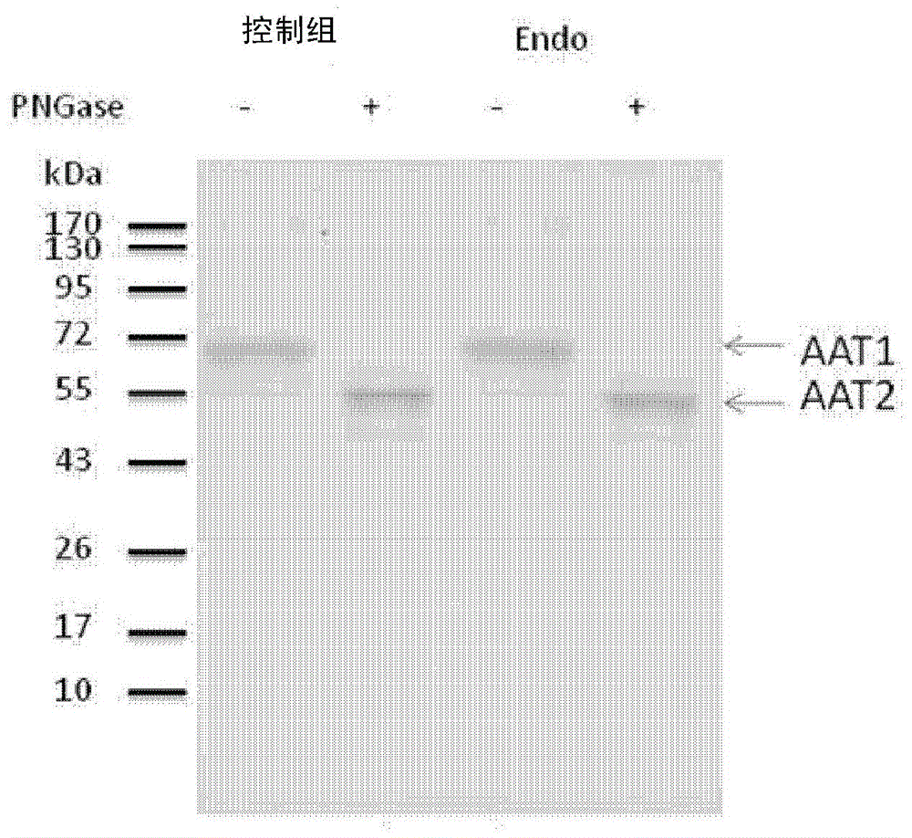
[0060] The protein contained in the above-mentioned rehydration buffer was treated with PNGase F to remove the N-glycan chains of the protein, and then the digested protein was id...
Embodiment 2
[0062] Example 2 Production of monoclonal antibodies
[0063] 2.1 Immunize animals with type A1-trypsin and measure the antibody titer
[0064] Mice were immunized with alpha 1-trypsin (A1AT, purchased from Sigma Inc), and a dose of 30 μg / animal was administered 2 to 6 times during a period of about 4 weeks. After the 2 booster doses, weekly blood samples were collected from the mice and the serum was immediately centrifuged. The separated serum was serially diluted, and then the antibody titer was measured by Western Blot.
[0065] 2.2 Preparation of antibody-producing cells
[0066] Select the animal of Example 2.1 with the desired antibody titer (i.e., in the western blotting method, the dilution concentration of the serum sample is 1:5,000, showing a positive reaction) for fusion Preparation from spleen cells, regional lymph nodes of immunized animals The antibody-producing cells were produced, and the antibody-producing cells were fused with the myeloma FO cell line ac...
Embodiment 3
[0073] Example 3 Detection of endometriosis using the monoclonal antibody of Example 2
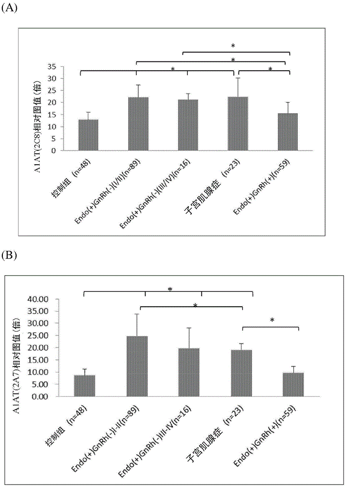
[0074] The specificity of monoclonal antibodies 2A7 and 2C8 in Example 2 was analyzed by immunoblotting method.
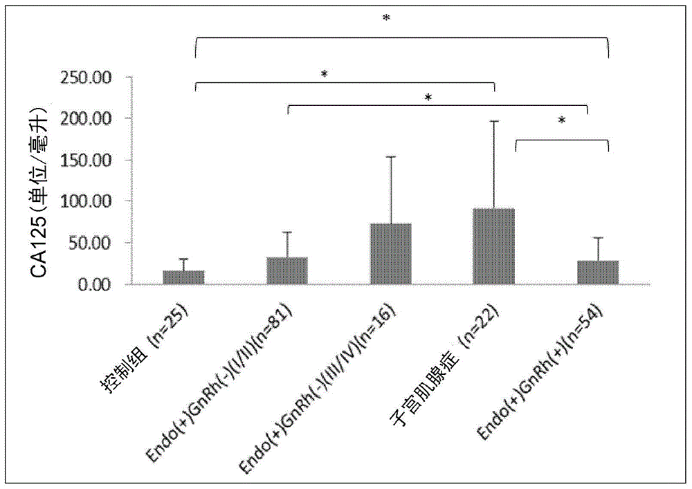
[0075] In this example, serum samples were obtained from 235 females who were informed and provided samples with their consent. These subjects were divided into five groups. Among them, the control group consisted of 48 pregnant women without endometriosis; the Endo(+)GnRh(-)I / II group consisted of 89 women with mild (or early) pelvic intrauterine Composed of women with ectopic membranes who did not receive gonadotropin-releasing hormone (GnRh) therapy; Endo(+)GnRh(-)III / IV group consisted of 16 women with severe pelvic endometriosis women who did not receive GnRH therapy; the adenomyosis group, which consisted of 23 women with adenomyosis; and finally, the Endo(+)GnRh(+) group, which consisted of 59 women with Consisting of women with pelvic endometriosis who had been treat...
PUM
| Property | Measurement | Unit |
|---|---|---|
| molecular weight | aaaaa | aaaaa |
| molecular weight | aaaaa | aaaaa |
Abstract
Description
Claims
Application Information
 - R&D
- Intellectual Property
- Life Sciences
- Materials
- Tech Scout
- Unparalleled Data Quality
- Higher Quality Content
- 60% Fewer Hallucinations
Browse by: Latest US Patents, China's latest patents, Technical Efficacy Thesaurus, Application Domain, Technology Topic, Popular Technical Reports.
© 2025 PatSnap. All rights reserved.Legal|Privacy policy|Modern Slavery Act Transparency Statement|Sitemap|About US| Contact US: help@patsnap.com