Intervertebral foramen mirror

A technique of intervertebral foraminal mirror and intervertebral disc, applied in the field of intervertebral foraminal mirror, can solve the problems of increasing the length of the instrument channel and the difficulty of instrument insertion, reducing the accuracy and stability of the surgical operation, increasing the difficulty of the operation and the risk of the operation, etc. Low surgical difficulty and risk, easy optical fiber patching, and shortening the impact distance

Inactive Publication Date: 2014-07-23
宋文生
View PDF5 Cites 4 Cited by
  • Summary
  • Abstract
  • Description
  • Claims
  • Application Information

AI Technical Summary

Problems solved by technology

However, the structural design of the currently used intervertebral foramina mirror is not reasonable enough. The cold light source interface is independent and vertically installed on the mirror stand. The near cold light source fiber is easy to collide with the body and hinder the rotation operation of the intervertebral foramen mirror; the angle between the eyepiece at the end of the mirror and the instrument channel is 30°-45°, and the projection distance of the eyepiece on the central axis of the instrument channel is long, and the eyepiece can control the operation of the instrument in the instrument channel The influence distance is large; the instrument channel is circular, and the difference between the outer diameter and the inner diameter of the instrument channel is small. After the longitudinal diameter of the intervertebral disc nucleus pulposus tissue is enlarged, it is difficult to pass through the instrument channel and the operation is complicated; In addition, the instrument channel ball water seal device is added, which increases the length of the instrument channel and the difficulty of instrument insertion; the slender endoscopic instruments and complicated surgical operations reduce their accuracy and stability, increase the difficulty and risk of the operation, and lead to steep learning curve

Method used


Image

  • Intervertebral foramen mirror

Examples


Embodiment Construction

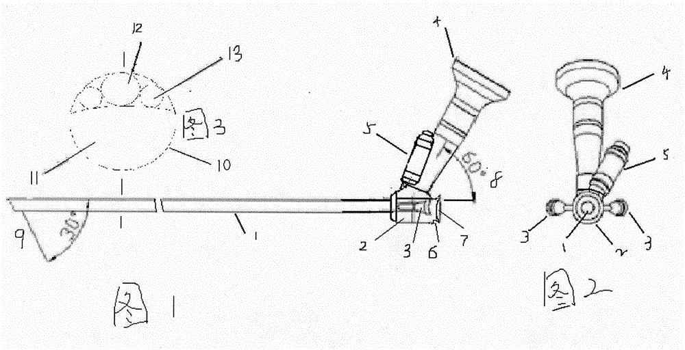
[0012] Intervertebral endoscope is a spinal endoscope that integrates lighting, observation, and surgical operation channels. Together with the corresponding supporting lighting, image system and endoscopic surgical instruments, it forms a spinal minimally invasive surgery system. During the operation, the intervertebral foramina mirror is connected with the liquid, cold light source, and image system. After the puncture is expanded and placed in the ideal working channel, the intervertebral foramina mirror is placed in front of the dural sac in the intervertebral foramen along the working channel. The instrument channel uses various surgical instruments under the microscope to remove the stained and prominent nucleus pulposus tissue, remove the bone, ablate the nucleus pulposus with radiofrequency electrodes, and seal the damaged annulus fibrosus.

[0013] The present invention will be further described below in conjunction with the accompanying drawings and embodiments.

[0...


PUM


Abstract

The invention relates to an intervertebral foramen mirror used in an intervertebral disc operation. The intervertebral disc comprises an instrument channel, an optical channel and two liquid channels, and is divided into a mirror platform part and a mirror tube part. The mirror platform is thicker than the mirror tube. An ocular provided with a cold light source loading interface is arranged above the mirror platform, and connected with the optical channel, and the included angle between the ocular and the optical channel is 120 degrees. The cold light source interface is located below the front lower end of the ocular, an included angle is formed between the cold light source interface and a front side plane, and the cold light source interface is nearly parallel to the ocular. The side wall of the mirror platform is provided with two liquid connectors to be connected with the corresponding liquid channels respectively. The tail of the mirror platform is provided with a water sealing cover groove, and is in a circular concave face. All working channel pipes are in a tumbler shape and are independent from each other to be arranged in the mirror tube in a sleeved and penetrating mode, the irregular oval instrument channel penetrates through the mirror tube part and the mirror platform part on the tumbler-shaped lower portion. The front end of the mirror tube is provided with an oblique face inclining downwards by 30 degrees. The intervertebral foramen mirror is short and high in definition, the length of the instrument under the mirror is small, inserting can be carried out easily, an operation is easy to carry out and high in precision and stability, 30-degree visual direction angle operation view and the operation space are large, the intervertebral foramen mirror conforms to the operation habit on an arthroscope, and the operation difficulty and risks are low.

Description

technical field [0001] The invention relates to a medical device, in particular a visible and minimally invasive surgical device for intervertebral disc herniation surgery, referred to as an intervertebral foramina mirror. Background technique [0002] The intervertebral foraminascope is a spinal endoscope that integrates lighting, observation, and surgical operation channels. The intervertebral foramina mirror enters the front of the dural sac in the intervertebral foramen at an angle of about 60° from the side and rear of the patient's body, displays it through the image system, and uses various surgical instruments under the instrument to remove the stained and prominent nucleus pulposus tissue, Bone removal, radiofrequency electrode ablation of the nucleus pulposus, and closure of the damaged annulus fibrosus. It is a minimally invasive spinal endoscope for the treatment of lumbar disc herniation with less trauma to patients and better results in similar operations. Ho...

Claims


Application Information

Patent Timeline
Patent Type & Authority Applications(China)
IPC IPC(8): A61B1/317
Inventor 宋文生
Owner 宋文生
Why Eureka
Try Eureka