Encephalic electrode individualization locating method based on multimode medical image data fusion

A medical imaging and data fusion technology, applied in medical science, surgery, diagnosis, etc., can solve problems such as registration error and image noise sensitivity, inability to guarantee EEG signals, rough intracranial electrode positioning, etc., and achieve high-precision individual The effect of optimized positioning and effective positioning

Inactive Publication Date: 2014-07-23
BEIJING NORMAL UNIVERSITY
View PDF2 Cites 35 Cited by
  • Summary
  • Abstract
  • Description
  • Claims
  • Application Information

AI Technical Summary

Problems solved by technology

Among the above two methods, the former uses a general standardized brain model instead of an individual's real brain model, and locates the intracranial electrodes in a relatively rough way, without considering the individualized positioning of the intracranial electrodes at all.
The latter is susceptible to the limitation of CT image data acquisition and the overall displacement of intracranial ele

Method used


Image

  • Encephalic electrode individualization locating method based on multimode medical image data fusion
  • Encephalic electrode individualization locating method based on multimode medical image data fusion
  • Encephalic electrode individualization locating method based on multimode medical image data fusion

Examples


Embodiment Construction

[0033] In order to make the object, technical solution and advantages of the present invention clearer, the present invention will be further described in detail below in conjunction with a specific embodiment and with reference to the accompanying drawings.

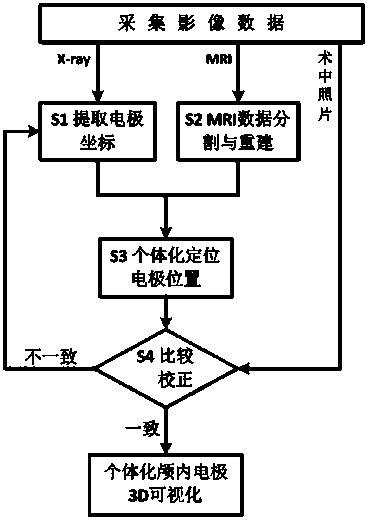
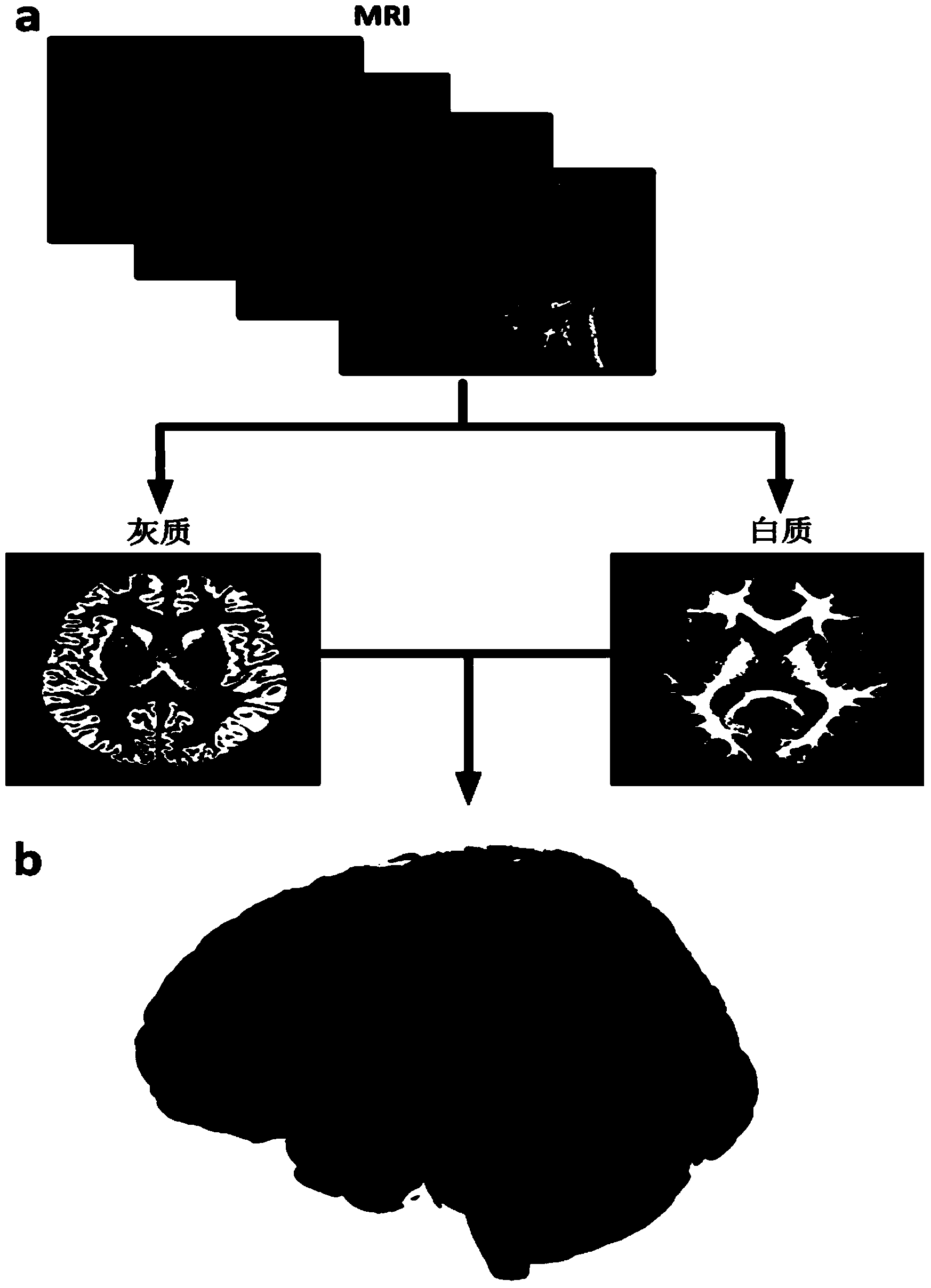
[0034] refer to figure 1 , a kind of intracranial electrode positioning method described in the present invention, based on multi-modal medical image data fusion to perform individualized intracranial electrode 3D visualization, the specific implementation steps are as follows:

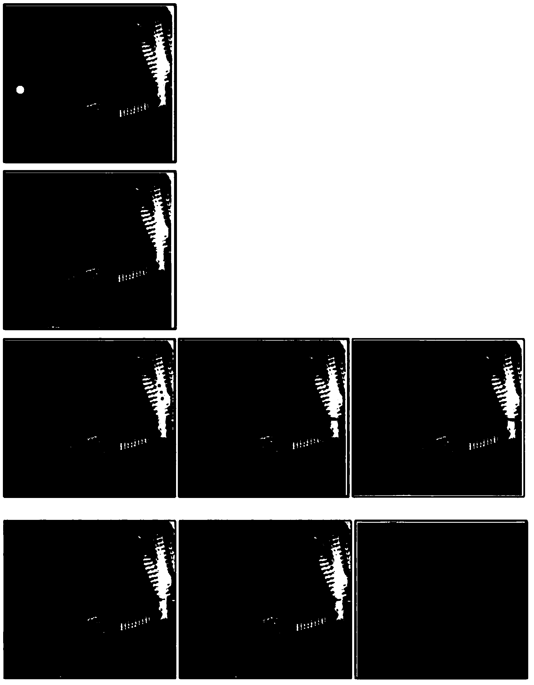
[0035] Step S1, image processing is performed on the plain X-ray film obtained from the X-ray examination of the brain, and electrode coordinates in the Talairach coordinate system are extracted.

[0036] Step 1.1, perform image processing on the X-ray plain film.

[0037] Since X-ray plain films are easily affected by detector noise during the acquisition process, in order to improve the image signal-to-noise ratio, the image needs to be prep...


PUM


Abstract

An encephalic electrode individualization locating method based on multimode medical image data fusion relates to the fields of medical instruments and medical image processing and neuroimaging. By means of multimode medical image data including magnetic resonance images, X-ray images and intranperative optical photos, the space position relation of an encephalic electrode and a brain tissue structure is comprehensively measured, a relation between the position of the encephalic electrode and a brain anatomical structure is established, and accurate and fast individualization locating of the encephalic electrode is achieved. The encephalic electrode individualization locating method based on multimode medical image data fusion can be used in the fields of cognitive neuroscience basic research, clinic neurosciences and the like, provide accurate brain space position information for an encephalic electroencephalogram signal, and enhance application value in the aspect of electrophysiological basis research.

Description

technical field [0001] The invention relates to the fields of medical equipment, medical image processing and neuroimaging, in particular to an intracranial electrode individualized positioning method based on multimodal medical image data fusion. Background technique [0002] With the development of digital technology and computer technology, scalp electroencephalogram (EEG) technology has made great achievements in the research and application of cognitive neuroscience and clinical disease auxiliary diagnosis with its extremely high time resolution. Due to the inherent deficiency of EEG's spatial positioning, currently EEG can only be used to measure the state of the brain's macroscopic brain regions. Magnetic resonance imaging (MRI) based on spatial encoding can provide complete brain structure information with its high spatial resolution, but its temporal resolution is relatively low. With the advancement of medical technology, intracranial electroencephalography (iEEG)...

Claims


Application Information

Patent Timeline
IPC IPC(8): A61B19/00
Inventor 李小俚胡振红
Owner BEIJING NORMAL UNIVERSITY
Why Eureka
Try Eureka