Endoscopic x-ray luminescence tomography device and method

A technology of luminescence tomography and imaging device, applied in the field of medical imaging

Active Publication Date: 2016-06-22
GUANGZHOU BLT INSTR & METER
View PDF7 Cites 3 Cited by
  • Summary
  • Abstract
  • Description
  • Claims
  • Application Information

AI Technical Summary

Problems solved by technology

However, due to the limitation of the penetration depth of the emitted near-infrared light, X

Method used


Image

  • Endoscopic x-ray luminescence tomography device and method
  • Endoscopic x-ray luminescence tomography device and method
  • Endoscopic x-ray luminescence tomography device and method

Examples


Embodiment Construction

[0064] The embodiments of the present invention will be described in detail below in conjunction with the accompanying drawings. The described examples are only intended to facilitate understanding of the present invention, and should not be construed as limiting the present invention.

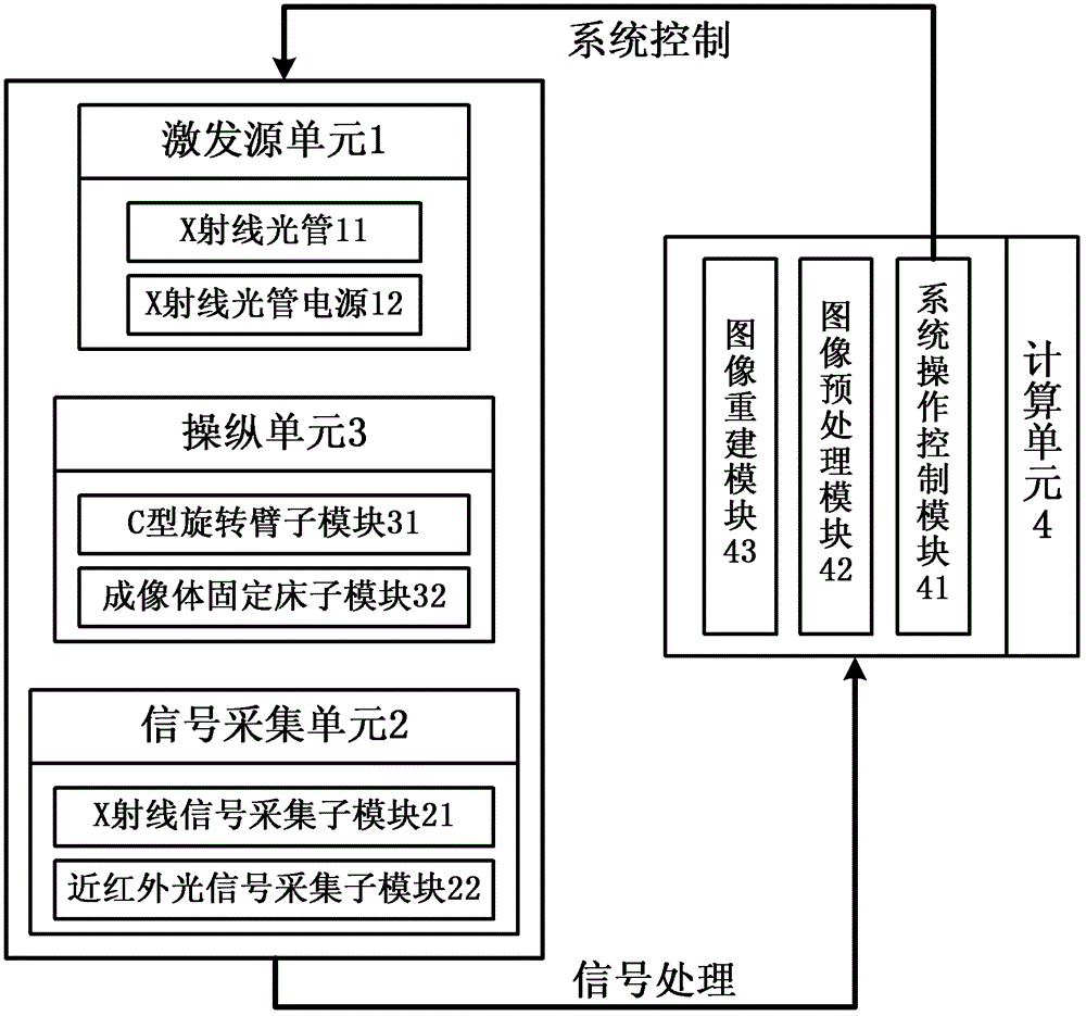
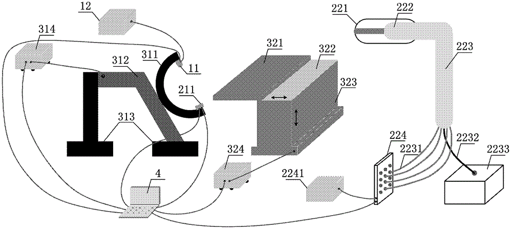
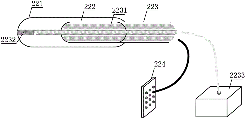
[0065] According to the embodiment of the present invention, the X-ray light tube is used as an external excitation source to excite the probe inside the object to be inspected, and at the same time, the miniature endoscopic probe is used as a detector to collect near-infrared light emitted by the probe after being excited. Then, the X-ray flat panel detector collinear with the X-ray light tube and the fixed bed of the imaging body is used to obtain the three-dimensional structure imaging information of the object under inspection. Based on the endoscopic X-ray luminescence tomography device and its imaging process, the physical model and forward light transmission model of endoscopic X-ray lum...


PUM


Abstract

An endoscopic X-ray luminescence tomography device, comprising: an excitation source unit (1), used to excite a probe inside an object under inspection from an external excitation source; a signal acquisition unit (2), used to collect the probe of the object under inspection The X-ray image and the near-infrared light signal image emitted after the probe is excited; the calculation unit (4) is used to preprocess the collected X-ray image and infrared light signal image, and to preprocess the X-ray image Sparse reconstruction is performed to obtain the structural information of the inspected object and the position information of the endoscopic probe in the signal acquisition unit inside the inspected object, and the preprocessed near-infrared light signal image is processed by using the obtained structural information and position information Perform optical three-dimensional reconstruction to obtain the position information and distribution information of the target inside the object under inspection.

Description

technical field [0001] The present invention relates to the technical field of medical imaging, and further relates to an endoscopic X-ray luminescence tomography device and an endoscopic X-ray luminescence tomography method. Background technique [0002] Because it can three-dimensionally reconstruct the spatial position and concentration distribution of fluorescent probes in organisms, fluorescence diffusion tomography (FDOT) has become a molecular imaging technology with great application potential. Among its potential applications, FDOT has been widely used in imaging small animals or superficial organs, such as brain and mammary gland. In this case, the fluorescent signal can penetrate the imaged object and be picked up by an external detection component. However, for the internal organs of the subject deeply buried in such as a large animal body or human body, due to the severe attenuation of fluorescence in the body, the FDOT technique cannot provide accurate images,...

Claims


Application Information

Patent Timeline
IPC IPC(8): A61B6/03
CPCA61B6/12A61B6/485A61B5/0086A61B6/032A61B6/5247
Inventor 梁继民陈雪利屈晓超朱守平陈多芳赵恒田捷
Owner GUANGZHOU BLT INSTR & METER
Why Eureka
Try Eureka