Reestablishing method of 4D-CT (Four Dimensional-Computed Tomography) different time phase sequence image

A 4D-CT, sequence image technology, applied in the field of image processing, can solve problems such as guiding placement error, affecting tumor boundary and effective judgment of motion law, etc., to achieve the effect of improving performance and avoiding injury

Inactive Publication Date: 2015-01-07
SHANDONG NORMAL UNIV
View PDF2 Cites 12 Cited by
  • Summary
  • Abstract
  • Description
  • Claims
  • Application Information

AI Technical Summary

Problems solved by technology

Although 4D-CT technology can effectively analyze the impact of respiratory movement on tumor target areas and organs at risk, 4D-CT is often acquired on the planned day, and the patient's tumor or other physical characteristics may change greatly on the treatment day. The best method is to obtain 3D-CT or CBCT images of the patient

Method used


Image

  • Reestablishing method of 4D-CT (Four Dimensional-Computed Tomography) different time phase sequence image
  • Reestablishing method of 4D-CT (Four Dimensional-Computed Tomography) different time phase sequence image
  • Reestablishing method of 4D-CT (Four Dimensional-Computed Tomography) different time phase sequence image

Examples


Embodiment Construction

[0048] Below in conjunction with accompanying drawing and embodiment the present invention will be further described:

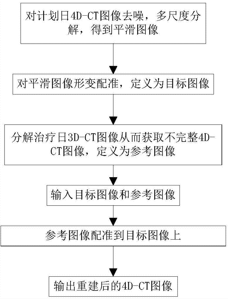
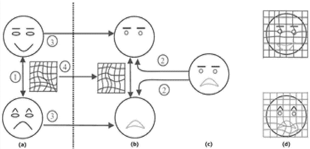
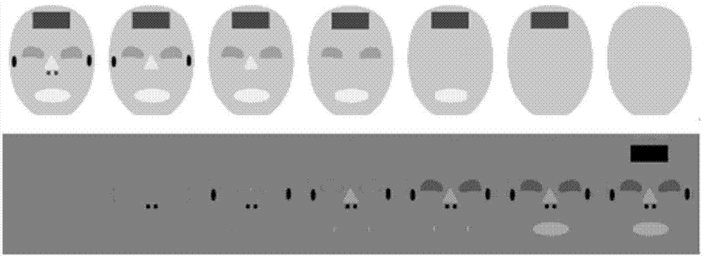
[0049] By constructing an edge-protected multi-scale space, the multi-scale decomposition of the 3D-CT images in the free-breathing state on the treatment day was carried out, and the phase attributes after the multi-scale decomposition of the images were determined by the similarity measure function. At the same time, combined with the B-spline-based free deformation mesh model to design a fully automatic registration scheme, it acts on the multi-scale 3D-CT on the radiotherapy day and the 4D-CT image on the planning day with different phase attributes, so as to reconstruct and describe the radiotherapy 4D-CT images on the day of treatment with tumor and organ-at-risk movements. Specific steps such as figure 1 Shown:

[0050] Step 1: Remove treatment bed artifacts from the 4D-CT images on the planned day, and remove noise or other oscillating components in t...


PUM


Abstract

The invention discloses a reestablishing method of a 4D-CT different time phase sequence image. The reestablishing method comprises the following steps: establishing an edge-protected multi-scale space, conducting multi-scale decomposition on a 3D-CT image on a therapeutic day under a free breathing state, determining an optimal time phase based on a maximal similarity measurement criterion, utilizing a free deformation model based on a spline B to realize deformation rectification of a 4D-CT different time phase image on a planned day, and reestablishing the 4D-CT image based on the 4D-CT image on the planned day and the 3D-CT image on the therapeutic day. The reestablishing method of the 4D-CT different time phase sequence image, disclosed by the invention, has the benefits that the 4D-CT image on the therapeutic day can be reestablished based on the 4D-CT image on the planned day and the 3D-CT image on the therapeutic day, so as to acquire the motion change law of tumor and endangered organ of a patient on the therapeutic day, and effectively avoid the damage to the body as the patient receives plenty of X-ray radiation again.

Description

technical field [0001] The invention relates to the field of image processing, in particular to a method for reconstructing 4D-CT images of different time-phase sequences. Background technique [0002] Clinical 4D-CT images bring large X-ray radiation to patients during the implementation process, and the impact of tumor and soft tissue movement on tumors and organs at risk, but based on the planned day (the physicist and clinician before the treatment of cancer patients for the patient Radiation treatment plan made using its 4D-CT image data) There is about a 1-2 week interval between the 4D-CT radiotherapy plan and the treatment day (during the radiotherapy of cancer patients) image data acquisition, and the tumor or other tissues may undergo major changes . If the 4D-CT is re-scanned every treatment day, the patient will again receive a large amount of X-ray radiation. [0003] Currently on treatment days, it is easy to obtain 3D-CT or CBCT (Cone beam CT) images, which ...

Claims


Application Information

Patent Timeline
IPC IPC(8): G06T11/00
Inventor 李登旺谭淑慧陈进琥李洪升尹勇
Owner SHANDONG NORMAL UNIV
Why Eureka
Try Eureka