Lung tissue model for biotoxicity detection and biotoxicity detection method

A technology of biological toxicity and detection method, applied in the field of biomedicine, can solve the problems of lack of three-dimensional characteristics of lung tissue in vivo, inability to accurately characterize the biological toxicity of nanoparticles, not being PM2.5, etc.

Active Publication Date: 2015-01-28
TSINGHUA UNIV
View PDF3 Cites 8 Cited by
  • Summary
  • Abstract
  • Description
  • Claims
  • Application Information

AI Technical Summary

Problems solved by technology

However, the traditional animal model involves issues such as immune accuracy, cost and ethics, and is not an ideal model for PM2.5 toxicity research
2D cell culture models lack the 3D characteristics of in vivo lung tissue, making it impossible to accurately characterize the biotoxicity of nanoparticles

Method used


Image

  • Lung tissue model for biotoxicity detection and biotoxicity detection method
  • Lung tissue model for biotoxicity detection and biotoxicity detection method
  • Lung tissue model for biotoxicity detection and biotoxicity detection method

Examples


Embodiment 1

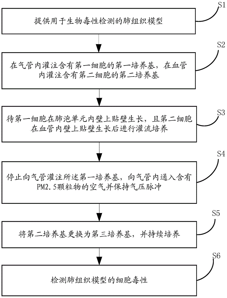
[0108] This implementation is to evaluate the pulmonary toxicity of PM2.5 by constructing a lung tissue model.

[0109] 1. Introduction of lung tissue model

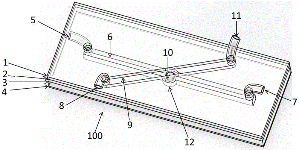
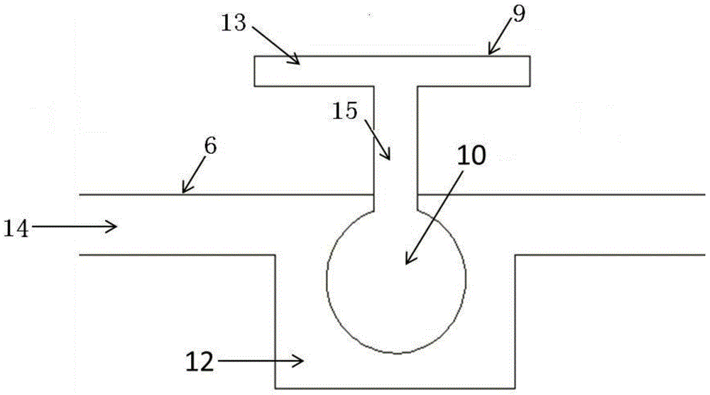
[0110] The lung tissue model in this embodiment is a chip structure. The lung chip device is mainly composed of three parts: trachea, blood vessel and alveolar unit. The trachea is a channel with a diameter of 200 microns, which is connected to the interior of the alveolar unit. Blood vessels are channels 200 microns in diameter that connect to the exterior of the alveolar unit. At the position connected to the alveolar unit is a cylindrical cavity structure with a diameter of 300 microns and a depth of 300 microns. Its inner wall is cultured with endothelial cells. The alveolar unit is a hollow spherical structure with a diameter of 200 microns and a membrane thickness of 10 microns. In addition, the alveolar unit membrane has a porous structure, and lung epithelial cells are cultured on the inner surface of the mem...

Embodiment 2

[0129] In this implementation, drug pulmonary toxicity evaluation is performed on the basis of the lung tissue model constructed in Example 1.

[0130] 1. Introduction of lung tissue model

[0131] The lung tissue model in this embodiment is the same as the lung tissue model in Embodiment 1, and will not be described in detail here.

[0132] 2. Manufacture and use of lung tissue model

[0133] In this example, 1. Chip manufacturing and culture process, 2. Material preparation process, 3. Chip printing process, 4. The steps of perfusing cells to construct a lung tissue model are the same as steps 1, 2, 3 and 4 in Example 1 , which will not be described in detail here.

[0134] 5. To detect the biological toxicity of the drug

[0135] Remove the medium supply of the gas channel, change it to normal air, and maintain a 0.3HZ air pressure pulse, so that the alveolar units can shrink and expand regularly; change the medium of the blood channel to EBM-2 and RPMI-1640 medium 1:1 ...

Embodiment 3

[0137] In this implementation, on the basis of the lung tissue model constructed in Example 1, pulmonary toxicity evaluation of inhaled drugs was carried out.

[0138] 1. Introduction of lung tissue model

[0139] The lung tissue model in this embodiment is the same as the lung tissue model in Embodiment 1, and will not be described in detail here.

[0140] 2. Manufacture and use of lung tissue model

[0141] In this example, 1. Chip manufacturing and culture process, 2. Material preparation process, 3. Chip printing process, 4. The steps of perfusing cells to construct a lung tissue model are the same as steps 1, 2, 3 and 4 in Example 1 , which will not be described in detail here.

[0142] 5. Test the biological toxicity of inhaled drugs

[0143] Remove the medium supply of the gas channel, change it to normal air, and maintain a 0.3HZ air pressure pulse, so that the alveolar units can shrink and expand regularly, and change the medium of the blood channel to EBM-2 and R...


PUM

PropertyMeasurementUnit
sizeaaaaaaaaaa

Abstract

The invention discloses a lung tissue model for biotoxicity detection and a biotoxicity detection method. The lung tissue model comprises a trachea, a blood vessel and an alveolus pulmonis unit, wherein an air passage is defined in the trachea, and an air inlet and an air outlet, which are communicated with the air passage respectively are formed at two ends of the trachea; a blood passage is defined inside the blood vessel, and a liquid inlet and a liquid outlet which are communicated with the blood passage respectively are formed at the two ends of the blood vessel, a cavity is defined in the alveolus pulmonis unit, an elastic ventilate membrane is formed on the wall of the alveolus pulmonis unit, the alveolus pulmonis unit is arranged in the blood passage, and the cavity is communicated with the air passage. By adopting the lung tissue model, an air exchanging function and a breathing strain effect of the lung blood in the body can be simulated; through planting cells in the alveolus pulmonis unit, the structure and function of the alveolus pulmonis unit in a body and inflammation reaction of immune cells can be simulated in the subsequent culture process.

Description

technical field [0001] The present invention relates to the field of biomedicine, in particular, to a lung tissue model for biotoxicity detection and a biotoxicity detection method, more specifically, to the in vitro bionic construction of the lung tissue model and the use of the model for environmental PM2.5 biological Toxicity and drug biotoxicity testing. Background technique [0002] According to the data released by the Beijing Municipal Environmental Protection Bureau, the number of good days in Beijing in 2013 was only 176 days, less than 50%. The cumulative number of days of heavy pollution at level 5 and level 6 reached 58 days. Air pollution (“smog”) has become a serious problem in China. Air pollution can cause acute and chronic respiratory diseases, heart disease, lung cancer and other diseases, seriously threatening human health. On March 25, 2014, the latest data released by the World Health Organization showed that the number of deaths due to air pollution ...

Claims


Application Information

Patent Timeline
Patent Type & Authority Applications(China)
IPC IPC(8): G01N33/15G01N33/48G09B23/28
Inventor 姚睿赵雨孙伟林峰
Owner TSINGHUA UNIV
Why Eureka
Try Eureka