Detection method for specific antibodies IgM of mycoplasma pneumonia and influenza viruses based on micro-fluidic chip

A technology of mycoplasma pneumoniae and microfluidic chip, which is applied in the field of biological analysis, can solve the problems of high cost of reagents and equipment, interference, etc., and achieve the effect of low price, small sample size, and quantitative reading results

Active Publication Date: 2015-02-18
THE NAT CENT FOR NANOSCI & TECH NCNST OF CHINA
View PDF9 Cites 11 Cited by
  • Summary
  • Abstract
  • Description
  • Claims
  • Application Information

AI Technical Summary

Problems solved by technology

Since this detection method involves biotin, there may be some interference due to the presence of biotin in human sample components. In addition, the cost of reagents and equipment used in fluorescence quantitative analysis is relatively high

Method used


Image

  • Detection method for specific antibodies IgM of mycoplasma pneumonia and influenza viruses based on micro-fluidic chip
  • Detection method for specific antibodies IgM of mycoplasma pneumonia and influenza viruses based on micro-fluidic chip
  • Detection method for specific antibodies IgM of mycoplasma pneumonia and influenza viruses based on micro-fluidic chip

Examples


Embodiment 1

[0063] Simultaneous detection of embodiment 1 mycoplasma pneumoniae and influenza virus specific antibody IgM

[0064] (1) Preparation of detection chip

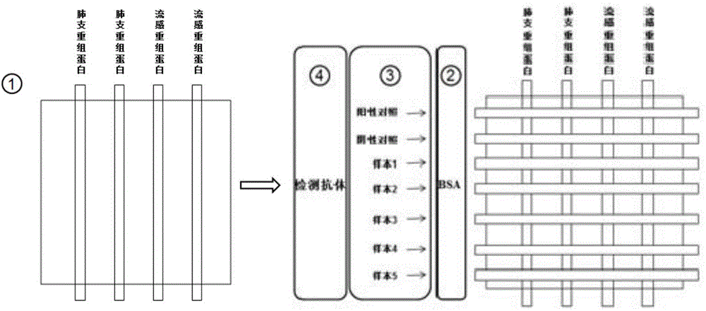
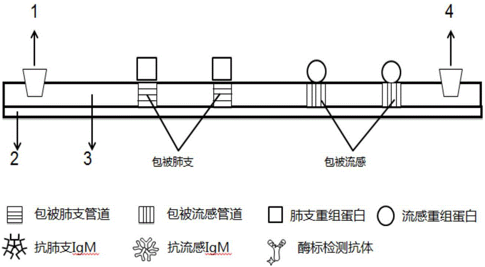
[0065] Take a multi-channel chip and seal it with the substrate, then take 1 μL of Mycoplasma pneumoniae recombinant protein stock solution and add it to PBS (PH7.2-7.4) buffer solution, dilute to the optimal coating concentration of 20 μg / mL, and mix well; the diluted protein The solution is passed into the two pipelines on the left; take 1 μL of influenza virus recombinant protein stock solution and add it to PBS (PH7.2-7.4) buffer solution, dilute to the optimal coating concentration of 15 μg / mL, and mix well; the diluted protein solution Pass into the two pipelines on the right; room temperature, coating for 30min.

[0066] Prepare 3% BSA blocking solution with PBS buffer. Remove the first chip, after air drying, paste the second chip, make the pipeline on the second chip perpendicular to the pipeline placement directi...

Embodiment 2

[0078] The detection of embodiment 2 Mycoplasma pneumoniae specific antibody IgM

[0079] (1) Preparation of detection chip

[0080] Take 1 μL of Mycoplasma pneumoniae recombinant protein and add it to PBS (pH7.2-7.4) buffer solution, dilute to the optimal coating concentration of 20 μg / mL, and mix well; take a multi-channel chip and seal it with the substrate; dilute the protein solution by The sample hole was connected to two pipelines, and was coated for 30 minutes at room temperature.

[0081] Prepare 3% BSA blocking solution with PBS buffer. Remove the first chip, after air drying, paste the second chip, make the pipeline on the second chip perpendicular to the pipeline placement direction of the first chip, and seal; then use a pipette to inject 20 μL BSA into the pipeline The blocking solution was kept at room temperature for 30 minutes; the blocking solution was taken out, dried, put into a sealed bag, and stored at 4°C to prepare a detection chip for Mycoplasma pneu...

Embodiment 3

[0093] The detection of embodiment 3 influenza virus specific antibody IgM

[0094] (1) Preparation of detection chip

[0095] Take 1 μL of influenza virus recombinant protein and add it to PBS (PH7.2-7.4) buffer solution, dilute to the optimal coating concentration of 15 μg / mL, mix well; take a multi-channel chip and seal it with the substrate; dilute the protein solution by The sample hole was connected to two pipelines, and was coated for 30 minutes at room temperature.

[0096] Prepare 3% BSA blocking solution with PBS buffer. Remove the first chip, after air drying, paste the second chip, make the pipeline on the second chip perpendicular to the pipeline placement direction of the first chip, and seal; then use a pipette to inject 20 μL BSA into the pipeline Blocking solution at room temperature for 30 minutes; take out the blocking solution, dry it, put it into a sealed bag, and store it at 4°C to prepare the detection chip.

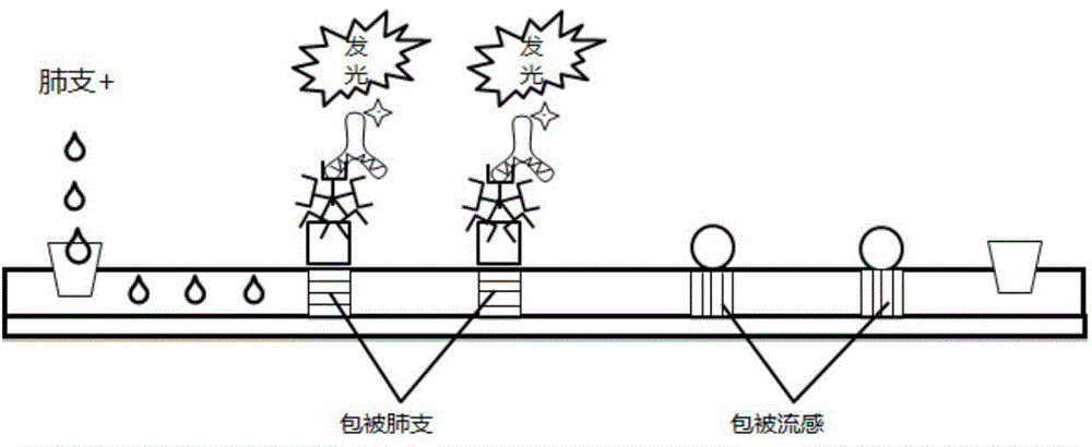
[0097] (2) Use the above-mentioned detect...


PUM

PropertyMeasurementUnit
lengthaaaaaaaaaa
diameteraaaaaaaaaa
heightaaaaaaaaaa

Abstract

The invention relates to a detection method for specific antibodies IgM of mycoplasma pneumonia and influenza viruses based on a micro-fluidic chip and application of the detection method. The method is characterized by taking the micro-fluidic chip with a plurality of pipelines as a reaction platform and detecting by using a chemical luminescent signal so as to independently or simultaneously detect the serum specific antibodies IgM of mycoplasma pneumonia and influenza viruses. As the micro-fluidic chip is combined with chemiluminiscence, the detection method integrates the advantages of rapidness, high efficiency and small sample amount of the micro-fluidic chip and high specificity of chemiluminiscence, also has the advantages of simplicity in operation, low cost, rapidness and the like, is a multi-target, real-time and high-sensitivity detection method and can be applied to rapid diagnosis of pneumonia and influenza.

Description

technical field [0001] The invention relates to the technical field of biological analysis, in particular to a method for detecting Mycoplasma pneumoniae and influenza virus, and in particular to a method for detecting the specific antibody IgM of Mycoplasma pneumoniae and influenza virus based on a microfluidic chip. Background technique [0002] Mycoplasma pneumoniae (Mycoplasma Pneumonia) is the pathogen of human mycoplasma pneumonia, which is characterized by paroxysmal irritating cough. In addition to the manifestations of the respiratory system, mycoplasma pneumonia can be accompanied by multi-system and multi-organ damage. The pathological changes of mycoplasma pneumonia are mainly interstitial pneumonia, sometimes accompanied by bronchial pneumonia, which is called primary atypical pneumonia. It is mainly transmitted by droplets, with an incubation period of 2 to 3 weeks. Some clinical symptoms are mild, or even asymptomatic. If there are, they are just general respi...

Claims


Application Information

Patent Timeline
Patent Type & Authority Applications(China)
IPC IPC(8): G01N33/569G01N33/545G01N21/76
CPCG01N33/545G01N33/56983G01N2333/11G01N2333/30
Inventor 蒋兴宇张晓青张伟曹丰晶陈翊平牛亚静沈海滢
Owner THE NAT CENT FOR NANOSCI & TECH NCNST OF CHINA
Why Eureka
Try Eureka