Freezing scanning electron microscope test method for nanoparticles in cosmetics

An electron microscope and detection method technology, applied in the field of testing, can solve problems such as deterioration, identification technology and lack of certification parties, agglomeration, etc., to achieve the effect of improving detection efficiency, shortening preparation time, and avoiding agglomeration and denaturation

Inactive Publication Date: 2015-02-18
CHINESE ACAD OF INSPECTION & QUARANTINE
View PDF2 Cites 6 Cited by
  • Summary
  • Abstract
  • Description
  • Claims
  • Application Information

AI Technical Summary

Problems solved by technology

Due to the extremely high physical and chemical activity of nanoparticles, serious agglomeration and deterioration will occur in such a complex extraction process, and the final product can no longer show the true state of these active ingredients in the product, resulting in subsequent qualitative analysis. and quantitative studies lack accuracy, and their corresponding risk assessments cannot be performed
This research status makes the identification technology and certification party of this kind of ingredients lack, and it is impossible to establish corresponding supporting testing institutions and rules and regulations

Method used


Image

  • Freezing scanning electron microscope test method for nanoparticles in cosmetics
  • Freezing scanning electron microscope test method for nanoparticles in cosmetics
  • Freezing scanning electron microscope test method for nanoparticles in cosmetics

Examples


Embodiment 1

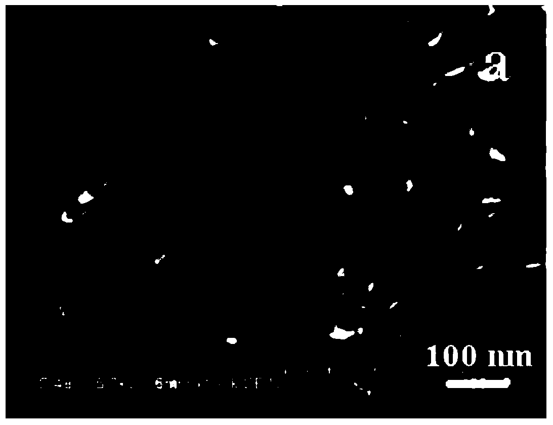
[0028] Example 1: Detection of Nanoparticles in Sunscreen

[0029] Take an appropriate amount of sunscreen sample and apply it in the groove of the sample holder with a thickness of 0.1mm to keep the sample holder in a horizontal state, put it into a freezer and freeze it for 20 minutes, transfer it to a sputtering instrument with a temperature of -112°C in the sample chamber, and coat it with a platinum film 60s, and then transferred to a scanning electron microscope with a sample chamber temperature of -106°C with a cryotransfer device for morphology testing, with a magnification of 30k, and EDS testing for the observed nanoparticles. From figure 1 It can be seen that there are needle-shaped nanoparticles in the sunscreen, with a length of about 80nm and a diameter of about 20nm. SEM observation showed that the nanoparticles were not agglomerated and distributed evenly.

[0030] As a comparison, the elemental composition of the sample was tested by traditional ICP (Inducti...

Embodiment 2

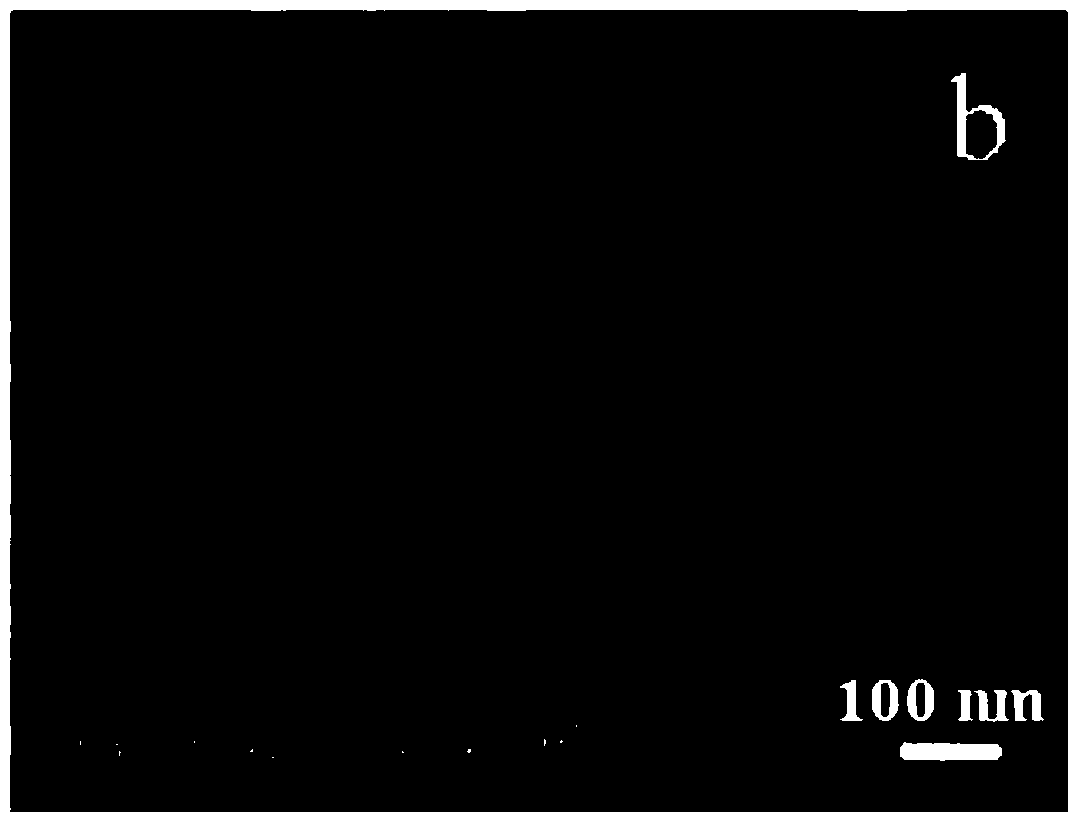
[0036] The detection of nanoparticle in embodiment 2 segregation cream

[0037] Take the isolation frost sample and apply it in the groove of the sample holder with a thickness of 0.1mm to keep the sample holder in a horizontal state, put it into the freezer and freeze it for 20 minutes, and transfer it to the sputtering instrument with the temperature of the sample chamber at -111°C with the frozen transfer device , gold-plated film for 60s, and then transferred to a scanning electron microscope with a sample chamber temperature of -105°C using a cryotransfer device for morphology testing, with a magnification of 100k, and EDS testing for the observed nanoparticles. From figure 2 It can be seen that there are spherical nanoparticles in sunscreen with a diameter of about 10nm. It can be seen from Table 2 that the composition of nanoparticles is SiO 2 , The quantitative detection results of EDS were consistent with the results of traditional ICP.

[0038] The composition an...

Embodiment 3

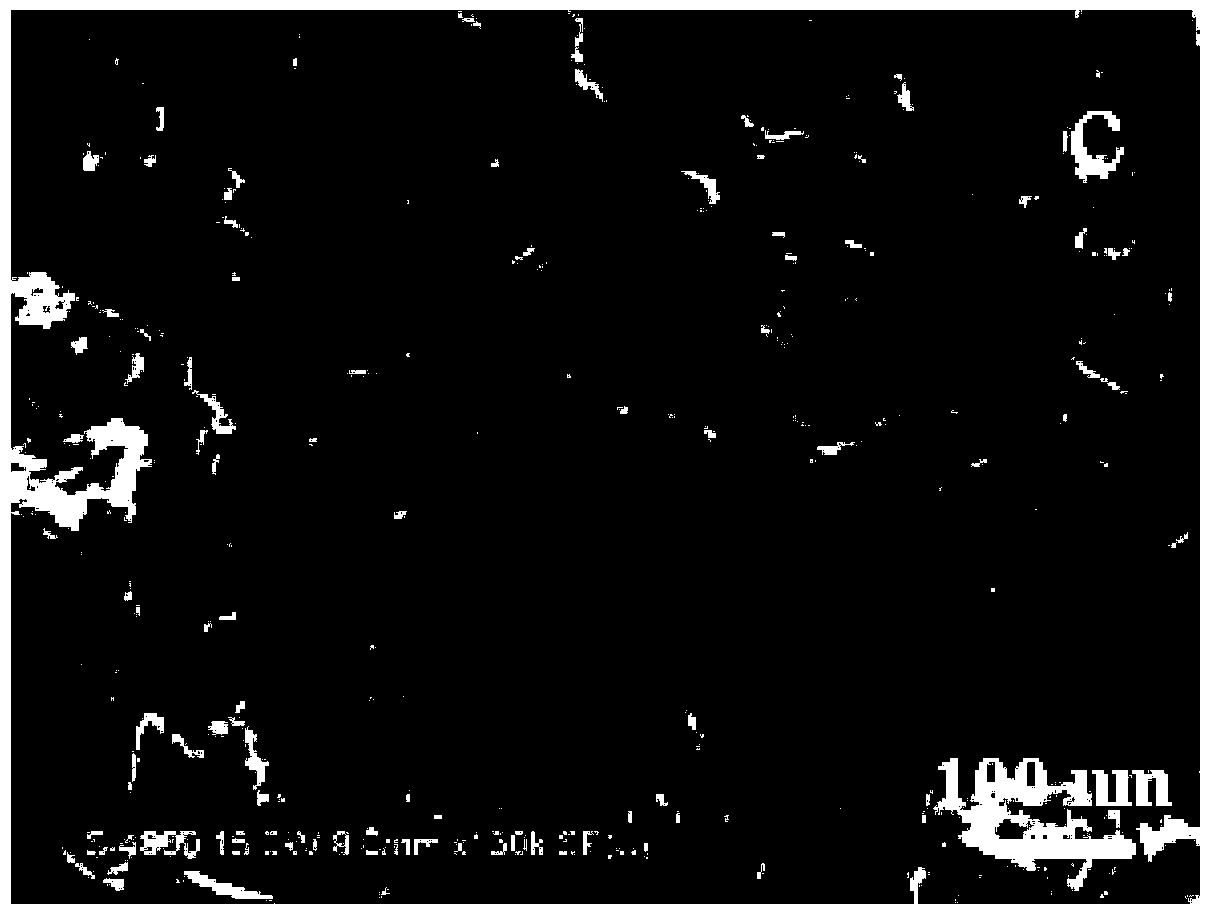
[0040] The detection of nanoparticle in embodiment 3 repairing face cream

[0041] Take an appropriate amount of repairing cream sample and apply it in the groove of the sample holder, the thickness is 0.1mm, so that the sample holder is kept in a horizontal state, put it into the freezer and freeze it for 20 minutes, transfer it to the sample chamber with a frozen transfer device at a temperature of -114°C for sputtering In the instrument, the film was coated for 60 s, and then transferred to a scanning electron microscope with a sample chamber temperature of -106°C by a cryotransfer device for morphology testing, with a magnification of 30k, and EDS testing was performed on the observed nanoparticles. From image 3 It can be seen that there are rod-shaped nanoparticles in the sunscreen, the length is about 60nm, and the diameter is about 30nm. It can be seen from Table 3 that the composition of nanoparticles is ZnO, and the quantitative detection results of EDS are consiste...


PUM


Abstract

The invention belongs to the field of testing and provides a freezing scanning electron microscope test method for nanoparticles in cosmetics. The method includes the steps of 1), placing cosmetic samples to be tested on a sample stand of a freezing scanning electron microscope, shifting the sample stand into a refrigerating unit, allowing the sample stand to be totally in liquid nitrogen, and performing refrigeration through the liquid nitrogen to have the samples frozen; 2), shifting the frozen samples to an ion sputtering unit for film coating; 3), after film coating, shifting the samples to the scanning electron microscope for morphology test. With the method, the samples to be studied are rapidly frozen under the condition of the liquid nitrogen so as to be perfectly kept in primitive forms, treated through a film coating technology and observed through the electron microscope; agglomeration and degeneration caused by pretreatment of nano components are avoided, preparation time for the real test samples is shortened, test efficiency is improved, true forms of the samples to be tested are completely restored, and accurate and reliable sample information is finally acquired.

Description

technical field [0001] The invention belongs to the field of testing, and in particular relates to a method for testing nanoparticles in cosmetics by using an electron optical device. Background technique [0002] Nanotechnology products have flourished in recent years and have had a huge impact on many fields such as biomedical research, electronics industry and chemical industry. With the continuous development of social economy, people pay more and more attention to beauty products, and the application of nanotechnology in cosmetics has attracted more and more attention. Nano ultrafine powder is extremely small in size and variable in shape, and has extremely high ductility and penetration. It can be used as an active substance with bactericidal and sunscreen effects, and can also be used as a carrier for other active ingredients. Research and application provide new avenues. [0003] At the same time, scholars and politicians around the world have expressed concern abo...

Claims


Application Information

Patent Timeline
Patent Type & Authority Applications(China)
IPC IPC(8): G01Q30/02G01Q30/20
Inventor 杨海峰李俊芳闫妍李亚辉席广成白桦
Owner CHINESE ACAD OF INSPECTION & QUARANTINE
Why Eureka
Try Eureka