Drug composition reducing in-vivo and in-vitro toxicity of nano drug delivery material and preparation method thereof

A technology of drug complexes and material bodies, applied in the biological field, can solve problems such as the weakening of autophagy ability, and achieve the effect of enhancing safety and reducing toxicity in vivo and in vitro

Inactive Publication Date: 2015-04-15
FUDAN UNIV
View PDF1 Cites 8 Cited by
  • Summary
  • Abstract
  • Description
  • Claims
  • Application Information

AI Technical Summary

Problems solved by technology

Most tumor cells (such as liver, pancreas, breast cancer, etc.) have different autophagy abilities before canceration, but their autophagy ability is weakened after canceration.

Method used


Image

  • Drug composition reducing in-vivo and in-vitro toxicity of nano drug delivery material and preparation method thereof
  • Drug composition reducing in-vivo and in-vitro toxicity of nano drug delivery material and preparation method thereof
  • Drug composition reducing in-vivo and in-vitro toxicity of nano drug delivery material and preparation method thereof

Examples


Embodiment 1

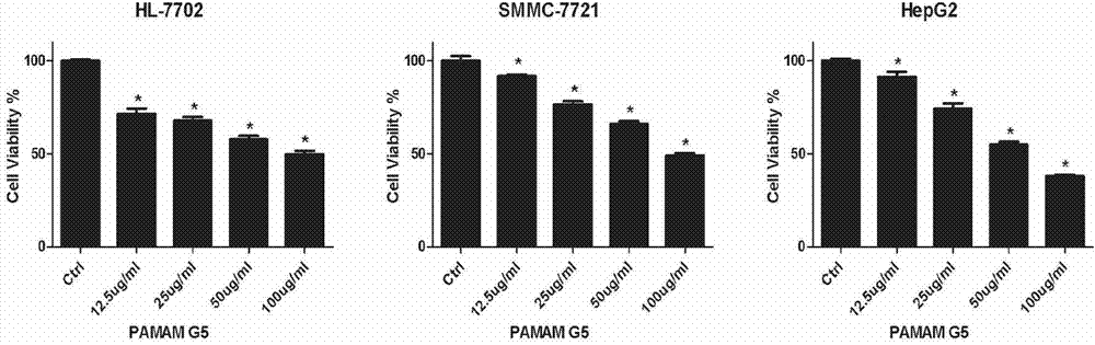
[0041] Example 1. Dendrimers are cytotoxic to human hepatocytes

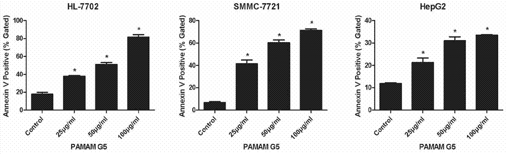
[0042] Normal human liver cells HL7702, liver cancer cells SMMC7721, and HepG2 were treated with different concentrations of PAMAM dendriemrs (12.5 μg / ml-100 μg / ml) for 24 hours, and the relative cell viability was measured by MTT method with untreated cells as negative control. The experimental results are as follows: figure 1 Shown; Annexin V / PI was used to stain cells, and flow cytometry was used to detect cell apoptosis. The experimental results were as follows figure 2 Shown; JC-1 was used to stain the next report, and flow cytometry was used to detect the collapse of mitochondrial membrane potential. The experimental results are as follows image 3 shown.

Embodiment 2

[0043] Example 2. Dendrimers Induce Autophagy in Human Hepatocytes

[0044] Human normal liver cells HL7702, liver cancer cells SMMC7721, and HepG2 were treated with 100 μg / ml dendrimers for 24 hours, then embedded in paraffin, sectioned, and stained, and the submicroscopic structure of the cells was observed under a transmission electron microscope. The results are as follows: Figure 4 As shown, there are a large number of typical double-membrane structure autophagosomes in the cells of the administration group, but not found in the control group.

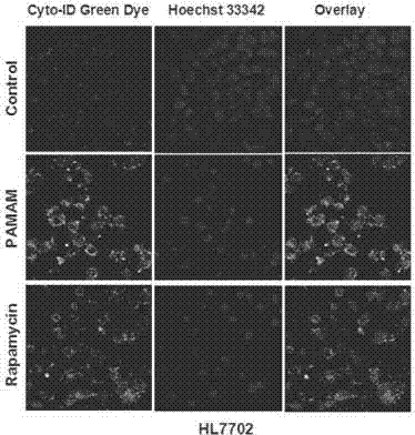
[0045] Normal human liver cells HL7702, liver cancer cells SMMC7721, and HepG2 were treated with 100 μg / ml dendrimers for 24 hours, stained with Cyto-ID autophagy detection fluorescent dye, and observed under a laser confocal microscope. The results are as follows: Figure 5 As shown, obvious green fluorescence can be observed in the cells of the administration group and the cells of the rapamycin effect group, while the green fl...

Embodiment 3

[0047] Example 3, 3-methyladenine inhibits autophagy and weakens hepatocyte growth inhibition caused by dendrimers

[0048] Human normal liver cells HL7702 were treated with different concentrations of PAMAM dendriemrs (12.5μg / ml-100μg / ml) for 24 hours, and 3-methyladenine was added to treat the cells 3 hours before administration, and the cell viability of each group was measured by MTT method after 24 hours . Experimental results such as Figure 7 As shown, 3-methyladenine pretreatment can significantly attenuate the growth inhibition of hepatocytes induced by dendrimers. The cells collected by centrifugation were washed once with PBS, and the cells were lysed with RIPA kits. After quantification, 20 μg of protein in each lane was electrophoresed and then transferred to PVDF membranes. Blocked with 5% skimmed milk for 1 h, LC3b and β-actin antibody, incubated at 4°C for 12h. After washing the membrane with TBST, the secondary antibody was added to incubate at room tempera...


PUM


Abstract

The invention belongs to the technical field of biology and relates to a drug composition reducing in-vivo and in-vitro toxicity of a nano drug delivery material and a preparation method thereof. In the invention, biological technologies are employed for reducing the in-vivo and in-vitro toxicity of the nano drug delivery material and the in-vivo and in-vitro toxicity of an autophagy-intervention nano material are controlled through the biological technologies, thereby reducing adverse side effects of the nano materials and improve safety of application of the nano materials. The invention particularly provides an autophagy-intervention composition which is composed of following substances including, but not limited to an autophagy-intervention drug (such as a compound or a polypeptide), an autophagy-related gene, an autophagy-related signal pathway protein blocking agent, with the nano material.

Description

Technical field: [0001] The invention belongs to the field of biotechnology, and relates to a method for regulating and controlling the toxicity of nano drug delivery materials in vivo and in vitro, in particular to a drug compound for reducing the in vivo and in vitro toxicity of nano drug delivery materials and its preparation method and application, more specifically, it provides a A kind of use of biotechnology to intervene in cell autophagy to regulate the in vivo and in vitro toxicity of nano drug delivery materials. Background technique: [0002] The prior art discloses that relevant nanomaterials mainly include nanoparticles, nanowires, nanofilms, nanotubes, and nanosolid materials. Since the size of such materials is in the intersection area between atomic clusters and macroscopic objects, nanomedical materials have the same characteristics as ordinary macroscopic materials. Therefore, it is widely used in drug delivery, disease diagnosis, tissue engineering, therap...

Claims


Application Information

Patent Timeline
Patent Type & Authority Applications(China)
IPC IPC(8): A61K45/00A61K48/00A61K49/00A61K47/34A61K47/04A61P39/02A61P35/00
Inventor 鞠佃文李玉彬曾贤钱晓璐王绍飞王子玉范佳君孙筠宋平冯美卿
Owner FUDAN UNIV
Why Eureka
Try Eureka