Sucking disc type external external-fixation uterus and oviduct radiography appliance

An external fixation, sucker-type technology, applied in the direction of drug devices, other medical devices, etc., can solve the problems of invading the uterine cavity, missed diagnosis of cervical canal lesions, pain and discomfort of the examinee, etc., to reduce pain, increase the contact area, The ideal effect of imaging

Inactive Publication Date: 2015-05-06
朱寒涛
View PDF15 Cites 2 Cited by
  • Summary
  • Abstract
  • Description
  • Claims
  • Application Information

AI Technical Summary

Problems solved by technology

All of the above-mentioned contrast devices have diagnostic and therapeutic defects such as balloon expansion of the cervical canal, causing pain and discomfort and invasion of the uterine cavity, and missed diagnosis of cervical canal lesions due to the occupation of the endocervical canal.

Method used


Image

  • Sucking disc type external external-fixation uterus and oviduct radiography appliance
  • Sucking disc type external external-fixation uterus and oviduct radiography appliance
  • Sucking disc type external external-fixation uterus and oviduct radiography appliance

Examples


Embodiment 1

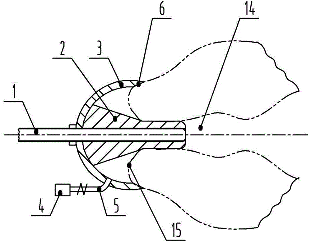
[0034] see figure 1 , 7 , in the figure, the suction cup type externally fixed hysterosalpingograph of the present invention includes a contrast agent injection tube 1, the inner end of which extends into the external os of the cervix 14, and its outer end is connected with a program-controlled drug infusion pump. The outer peripheral surface of the inner end of the drug injection tube is fixedly fitted with a conical head 2 and a suction cup from the inside to the outside in sequence. Matching with the joint part of the suction cup, the contrast device also includes a vacuum pumping mechanism 4 , and the vacuum pumping mechanism communicates with the inner chamber of the suction cup through a flexible tube 5 .

[0035] The opening edge of the inner end of the suction cup is provided with a bevel 6 that matches and contacts the outer end of the uterus. The groove surface is a rough surface (such as a frosted surface), a corrugated surface or a transverse thread surface. Bot...

Embodiment 2

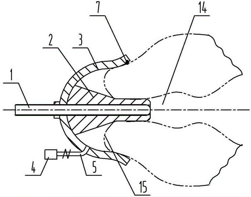
[0041] see figure 2 , in the figure, this embodiment is similar in structure to Embodiment 1, and the structurally identical parts will not be repeated here. Bell mouth 7; the bell mouth surface is a rough surface (such as a frosted surface), a corrugated surface or a transverse thread surface.

Embodiment 3

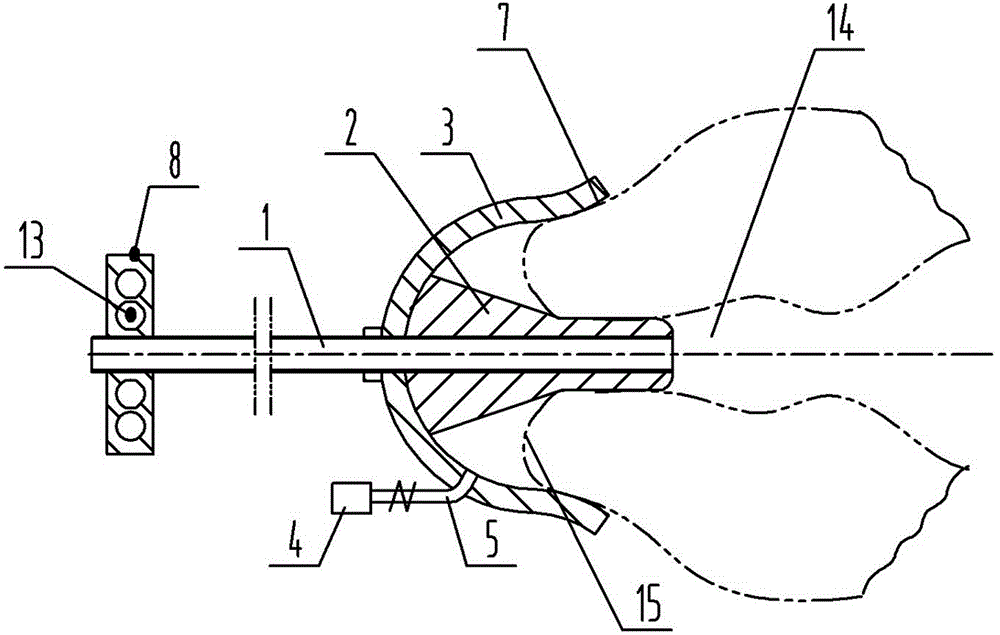
[0043] see image 3 , in the figure, the structure of this embodiment is similar to that of Embodiment 2, and the structurally identical parts will not be repeated here. Plate type 8, and the handle is provided with finger jack 13. The setting of the handle is convenient for the operator to operate at a place away from the workbench, so as to avoid radiation and help protect the staff.


PUM


Abstract

The invention belongs to the technical field of medical obstetrical and gynecological examination and particularly relates to a sucking disc type external external-fixation uterus and oviduct radiography appliance. The sucking disc type external external-fixation uterus and oviduct radiography appliance comprises a contrast medium injection tube and further comprises a vacuum-pumping mechanism, wherein the inner end of the injection tube extends into an external cervical orifice and the outer end of the injection tube is connected with a program-controlled medicine infusion pump; a tapered head and a sucking disc are fixedly arranged on an outer peripheral surface of the inner end of the contrast medium injection tube from inside to outside in sequence in a sleeving mode; the tapered head comprises a cylindrical section which is matched with the external cervical orifice in inner diameter and a tapered section which is matched and positioned with the external cervical orifice; the inner end of the sucking disc is open and buckled at the outer end of a uterus; joint parts of the tapered head and the sucking disc are coincided and matched; the vacuum-pumping mechanism is communicated with an inner cavity of the sucking disc through a hose. The sucking disc type external external-fixation uterus and oviduct radiography appliance is relatively firm in fixation, relatively good in stability, free of needs of implantation, complete in examination and ideal in contrast effect, and does not cause pains and discomfort to a patient, so that cross infection and cervical impairment can be avoided, x-ray radiation is avoided, and operation staff are protected in a facilitated manner.

Description

technical field [0001] The invention belongs to the technical field of medical obstetrics and gynecology inspection, in particular to a suction cup type externally fixed hysterosalpingograph. Background technique [0002] Infertility is a common disease in gynecology. There are many reasons for infertility, among which lumen obstruction caused by salpingitis is the most common. Clinical use of transvaginal hysterosalpingography to check the patency of the fallopian tubes. At present, the clinically used appliances in China mainly include: 1. Fallopian tube ventilator: made of metal, with a hard piston at the front end. Two people are required to operate during radiography, which is inconvenient to operate, easy to leak medicine, and easy to cause cervical injury to patients; 2. Fallopian tube Imaging off-line device: on the basis of the above-mentioned fallopian tube ventilator, a metal iron seat is added to fix the cervical forceps and imaging tube, so that the staff can ...

Claims


Application Information

Patent Timeline
Patent Type & Authority Applications(China)
IPC IPC(8): A61M31/00
Inventor 朱寒涛乔新荣
Owner 朱寒涛
Why Eureka
Try Eureka