Left Ventricular Volume Reducing Device

A technology of left ventricle and main body, which is applied in the field of left ventricle volume reduction device, can solve the problems that the position cannot reach the optimal position, the doctor’s operation requirements are relatively high, and the success rate of surgery is low, so as to reduce the risk of surgery and complications, and reduce the risk of surgery. Risk, effect of simplified operation

Active Publication Date: 2018-04-10
SHENZHEN KYD BIOMEDICAL TECH CO LTD
View PDF5 Cites 0 Cited by
  • Summary
  • Abstract
  • Description
  • Claims
  • Application Information

AI Technical Summary

Problems solved by technology

However, if the device is placed in a bad position at one time, it cannot be recovered for repeated adjustments
As a result, the doctor's operation requirements are relatively high, so that the position of the instrument placement cannot reach the optimal position, and the success rate of the operation is low

Method used


Image

  • Left Ventricular Volume Reducing Device
  • Left Ventricular Volume Reducing Device
  • Left Ventricular Volume Reducing Device

Examples


Embodiment 1

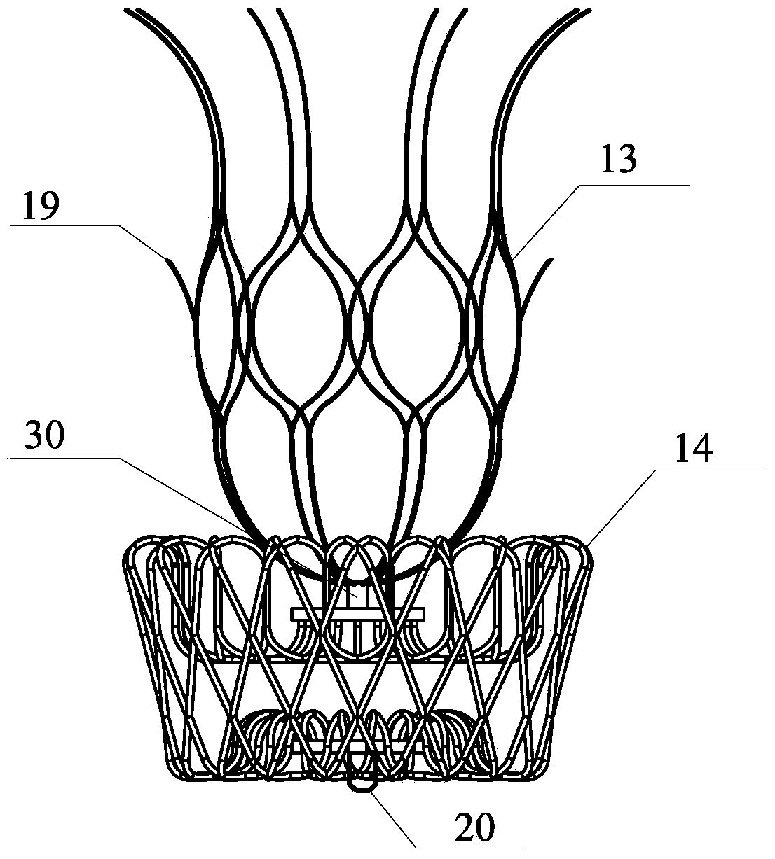
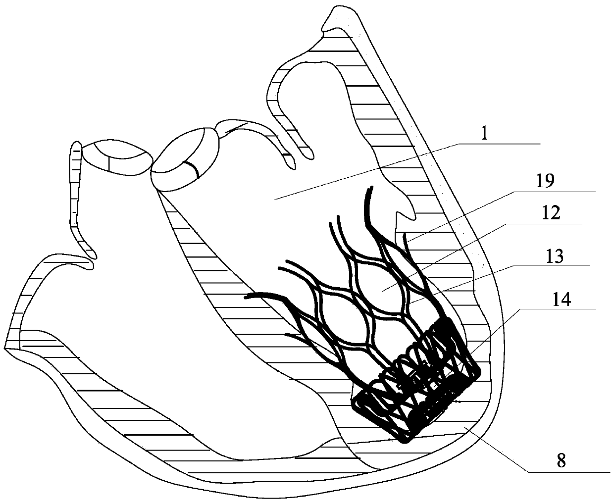
[0039] like figure 1 , 2 As shown, the left ventricular volume reduction device of the present invention adopts a two-stage design, which are the main body 13 and the base 14 respectively. The main body 13 is integrally formed by laser cutting of a nickel-titanium alloy tube, and the nickel-titanium alloy tube used has anti-magnetic properties and does not affect nuclear magnetic resonance imaging. Its cage-like structure has excellent mechanical stability, is not easy to deform, break and fall apart, and maintains its structural integrity. like figure 2 As shown, the left ventricular volume reduction device involved in the present invention can be placed in the left ventricle 1 through the aorta or through the apex, the base 14 is located at the position of the left apex 8, and the main body 13 is attached to the outer wall of the left ventricle 1 and the interventricular septum 3 Placed above the base 14, isolate the inactivated myocardium below the left ventricle 1, red...

Embodiment 2

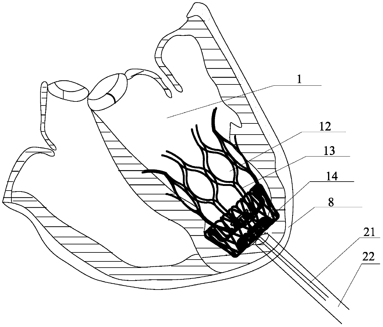
[0042] like Figure 5 , 6 , 7a, 7b, and 7c, this embodiment adopts a three-stage design, which are the main body 16, the base 17 and the blocking disc 18 respectively. The main body 16 is integrally formed by laser cutting of a nickel-titanium alloy tube, and the nickel-titanium alloy tube used has anti-magnetic properties and does not affect nuclear magnetic resonance imaging. Its cage-like structure has excellent mechanical stability, is not easy to deform, break and fall apart, and maintains its structural integrity. The left ventricular volume reduction device involved in the present invention can be placed in the left ventricle 1 through the aorta or through the apex, the occlusion disc 18 is placed outside the myocardium of the apex of the left ventricle, the base 17 is located at the position of the left apex 8, and the main body 16 is placed above the base against the outer wall of the left ventricle 1 and the interventricular septum 3 to isolate the inactivated myoc...

Embodiment 3

[0045] like Figure 8 and Figure 9 As shown, the left ventricular volume reduction device of the present invention adopts a two-stage design, which are the main body 13 and the base 14 respectively. The main body 13 is integrally formed by laser cutting of a nickel-titanium alloy tube, and the nickel-titanium alloy tube used has anti-magnetic properties and does not affect nuclear magnetic resonance imaging. Its cage-like structure has excellent mechanical stability, is not easy to deform, break and fall apart, and maintains its structural integrity. like figure 2 As shown, the left ventricular volume reduction device involved in the present invention can be placed in the left ventricle 1 through the aorta or through the apex, the base 14 is located at the position of the left apex 8, and the main body 13 is attached to the outer wall of the left ventricle 1 and the interventricular septum 3 Placed above the base, it isolates the inactivated myocardium below the left vent...


PUM


Abstract

The invention discloses a left ventricular volume reduction device, which adopts a two-stage design and includes: a base and a main body, and the shape of the base is a cage-like structure. The main body is formed by integral laser cutting of a nickel-titanium alloy tube and is in the shape of multiple The design of the grid is a layered grid, and the grid is a closed-loop structure design. The top of the main body is also provided with an upward-inclined anchor spine. When placed, the anchor spine can penetrate into the myocardium of the ventricular wall to anchor the left ventricle. The function of the volume reducing device is that the lower end of the main body is connected to a tubular structure, and the tubular structure is connected to the base. The base is woven into a braided mesh structure through nickel titanium wire, and is heat-treated into a disk shape. The invention is a repositionable, fixed and stable left ventricular volume reduction device.

Description

technical field [0001] The invention relates to the technical field of left ventricular volume reduction devices, in particular to a repositionable, fixed and stable left ventricular volume reduction device. Background technique [0002] Heart failure is a common debilitating and potentially fatal condition in which the heart cannot provide enough blood flow to meet the body's needs. Symptoms of heart failure that negatively affect quality of life include shortness of breath, persistent coughing or wheezing, excessive fluid buildup in body tissues (edema), fatigue, loss of appetite or nausea, slowed thinking, and rapid heart rate. In recent years, the prevalence of heart failure has increased year by year and has become a serious public health problem. It is reported that 17 million people die of cardiovascular disease in the world every year, and more than half of them die of acute myocardial infarction. Even if patients survive, they are prone to develop chronic diseases. ...

Claims


Application Information

Patent Timeline
Patent Type & Authority Patents(China)
IPC IPC(8): A61B17/00A61B17/12
CPCA61B17/0057A61B17/12022A61B17/12122A61B17/12172A61B2017/1205A61B2017/00004A61B2017/00243A61B2017/00597A61B2017/00632A61B2017/00668A61F2/2442
Inventor 陈奕龙黄伟
Owner SHENZHEN KYD BIOMEDICAL TECH CO LTD
Why Eureka
Try Eureka