A blood cell mechanical stress deformation pulse laser synchronous microscopic imaging observation device

A microscopic imaging and pulsed laser technology, which is applied in application, medical science, surgery, etc., can solve the problems of mechanical damage research of red blood cells, high cost of laser diffractometer, lack of blood damage model, etc., and achieves strong deformation observation flexibility, Easy to observe, high uniformity effect

Inactive Publication Date: 2017-06-13
SUZHOU UNIV
View PDF6 Cites 0 Cited by
  • Summary
  • Abstract
  • Description
  • Claims
  • Application Information

AI Technical Summary

Problems solved by technology

However, its research direction is limited to the hemodynamic properties of the artificial heart, and does not study the mechanical damage of red blood cells.
Automatic laser diffractometer is a device for measuring the deformability, orientation, quantity, diameter, surface area, etc. of red blood cells in blood, so as to conduct research on blood rheological characteristics. It has a broad application background, but lacks an effective blood damage model. Obtain a series of experimental data with high reliability, and the cost of laser diffractometer is relatively expensive
[0006] So far, the device used to observe the mechanical stress and deformation of blood cells still needs to be perfected. There is no method that can not only meet the needs of blood mechanical injury experiments, but also obtain a blood injury model with high reliability, and can also visualize and observe the characteristics of the interstitial flow field in real time. , The device for analyzing the distribution of shear stress

Method used


Image

  • A blood cell mechanical stress deformation pulse laser synchronous microscopic imaging observation device
  • A blood cell mechanical stress deformation pulse laser synchronous microscopic imaging observation device
  • A blood cell mechanical stress deformation pulse laser synchronous microscopic imaging observation device

Examples


Embodiment

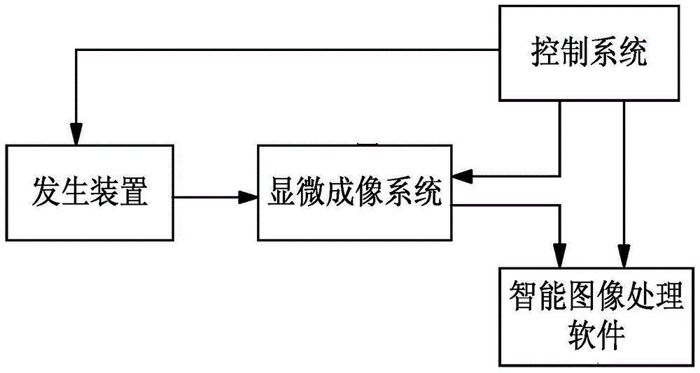
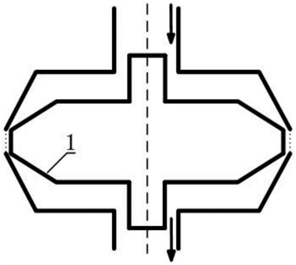
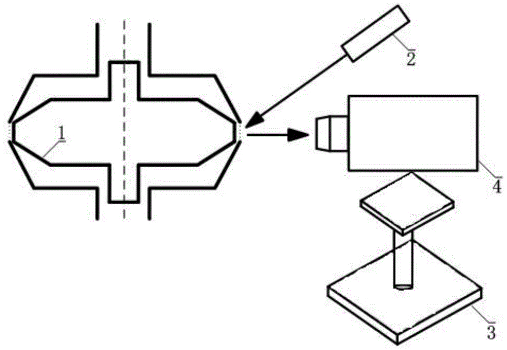
[0023] Example: such as figure 1 As shown, a blood cell mechanical stress deformation observation device structure includes a generating device, a microscopic imaging system, a control system and a data analysis system, blood cells are placed in the generating device, and an observation device is set on the generating device gap, the microscopic imaging system is located on the side of the generating device close to the observation gap, and the generating device, the microscopic imaging system and the data analysis system are all electrically communicated with the control system. The generating device is used to generate a shear field to deform the blood cells. Micro imaging system, control system and data analysis system make up the observation device. The control system controls the injection of the blood medium in the generating device to generate a shear field, controls the microscopic imaging system to capture images of marked blood cells, controls the data analysis syst...


PUM


Abstract

The invention discloses a blood cell mechanical stress deformation pulsed laser synchronous microscopic imaging observation device, which includes a generating device, a microscopic imaging system, a control system and a data analysis system. Blood cells are placed in the generating device, and the An observation gap is opened on the generating device, and the microscopic imaging system is located on a side of the generating device close to the observation gap, and the generating device, the microscopic imaging system and the data analysis system are all connected with the control System electrical connection. A shear field is generated by the generator to deform the blood cells. The control system controls the injection of the blood medium of the launch device and the generation of the shear field, controls the microscopic imaging system to capture images of the marked blood cells, controls the data analysis system to automatically extract the red blood cell deformation parameters in real time, and performs data and image analysis and processing . Simple operation, high sensitivity and convenient observation.

Description

technical field [0001] The invention relates to an observation device for mechanical stress deformation of blood cells, in particular to a pulse laser synchronous microscopic imaging observation device for mechanical stress deformation of blood cells. Background technique [0002] Since the advent of cardiovascular artificial organs, related research and applications have developed rapidly, and many of them have been used clinically. Nevertheless, the law of blood mechanical damage in artificial organs (especially the thrombus and hemolysis caused by red blood cell damage) has always been an unresolved problem, which seriously hinders the clinical promotion and use of medical devices such as cardiovascular artificial organs. Therefore, the research on the law of blood mechanical damage is a core content of the development of artificial organs and other medical devices. Designing an intuitive and accurate blood injury experimental device can provide experimental means for th...

Claims


Application Information

Patent Timeline
Patent Type & Authority Patents(China)
IPC IPC(8): A61B90/00
Inventor 徐博翎杨磊尹成科孙宏健陈霄史凤燕张心宇尤志强
Owner SUZHOU UNIV
Why Eureka
Try Eureka