
Fluorescent RT-PCR detection reagent of nipah virus M gene as well as preparation method and application of fluorescent RT-PCR detection reagent

A technology of RT-PCR, Nipah virus, applied in the field of biology

Inactive Publication Date: 2015-07-15
ANIMAL & PLANT & FOOD INSPECTION CENT OF TIANJIN ENTRY EXIT INSPECTION & QUARANTINE BUREAU
View PDF2 Cites 2 Cited by
  • Summary
  • Abstract
  • Description
  • Claims
  • Application Information

AI Technical Summary

Problems solved by technology

At present, the laboratory methods for Nipah virus detection mainly include virus isolation and identification, fluorescent antibody test for detection of pathogens and RT-PCR method for detection of viral nucleic acid (Daniels P, Ksiazek T, Eaton B, Laboratory diagnosis of Nipah and Hendra virus infections Microbes Infect, 2001.3: p.289-295.), and for detecting the fluorescent RT-PCR detection reagent of Nipah virus M gene and its preparation method and application, there is no report yet

Method used

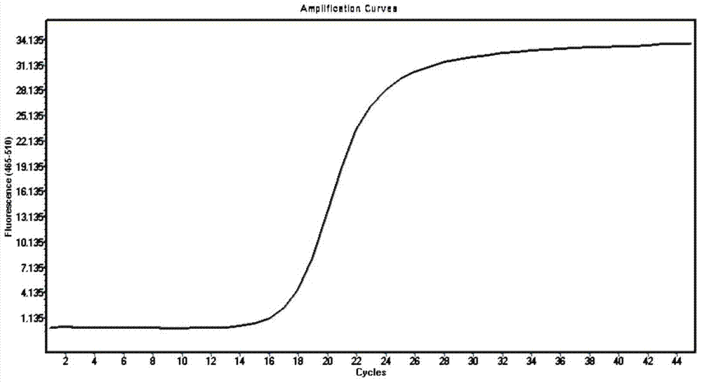
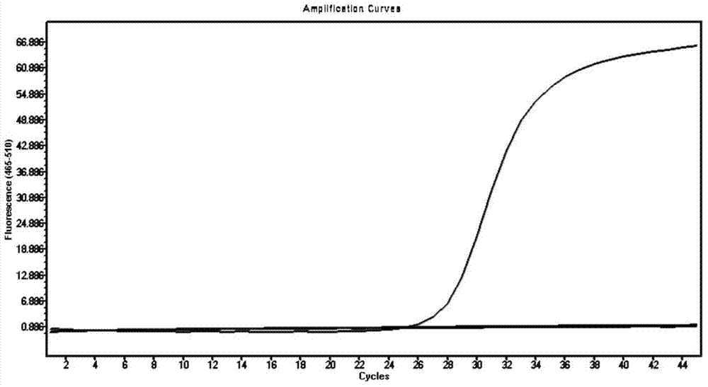
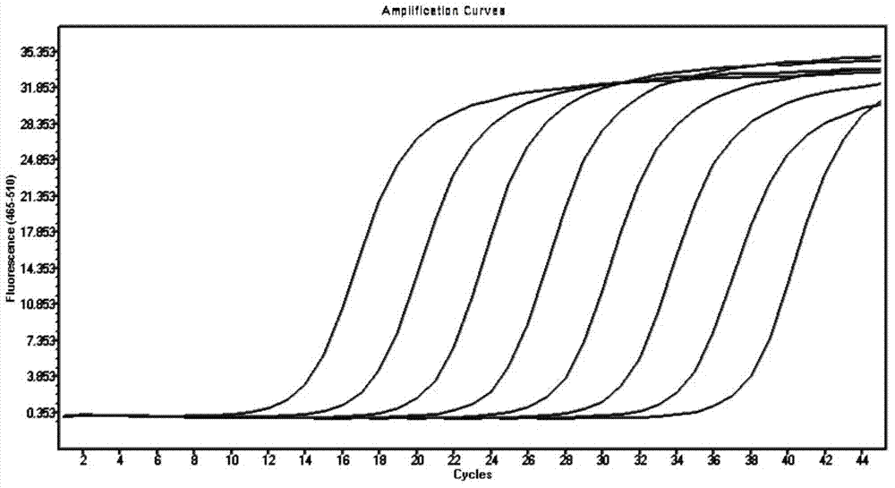

Image

  • Fluorescent RT-PCR detection reagent of nipah virus M gene as well as preparation method and application of fluorescent RT-PCR detection reagent
  • Fluorescent RT-PCR detection reagent of nipah virus M gene as well as preparation method and application of fluorescent RT-PCR detection reagent
  • Fluorescent RT-PCR detection reagent of nipah virus M gene as well as preparation method and application of fluorescent RT-PCR detection reagent

Examples


preparation example Construction

[0032] The fluorescent RT-PCR detection reagent of Nipah virus M gene and its preparation method and application of the present invention comprise the following steps.

[0033] The first step is to prepare the positive control substance of Nipah virus M gene, and the process includes:

[0034] (1) In the GenBank database, the M gene sequence was compared by BLAST, and a conserved sequence was selected for the preparation of the M gene positive control, as shown in SEQ ID NO.4.

[0035] (2) Design and synthesize 4 primers based on the sequence of SEQ ID NO.4, the primer sequences are as follows:

[0036] NIPAH-M-F1:5'-CCTGGAACAACAGTTGTGAGATCAGCCGAGTAGCAGCTGTGTTGCAGCCTTCTGTTCC-3'

[0037] NIPAH-M-R1:5’-GGAACAGAAGGCTGCAACACAGCTGCTACTCGGCTGATCTCACAACTGTTGTTCCAGG-3’

[0038] NIPAH-M-F2:5'-GATGGACATCAATCCTTGGCTCAACAGATTGACCTGGAACAACAGTTGTGAGATC-3'

[0039] NIPAH-M-R2: 5'-GAAGACATCATCATAGATCATGAACTCTCTTGGAACAGAAGGCTGCAACACAGCTG-3';

[0040] (3) two complementary primers are used as...

specific Embodiment approach

[0068] The present invention will be further described in detail below in conjunction with the accompanying drawings and specific embodiments.

example 1

[0069] The preparation of example 1 Nipah virus M gene positive control

[0070] 1. Selection of reference gene sequence

[0071] BLAST was performed on the Nipah virus M gene sequence (GenBank accession number: AJ564621.1), and a conservative and suitable sequence for designing fluorescent RT-PCR primers and probes was selected as a reference sequence for preparing Nipah virus M gene positive control (RNA) , as shown in SEQ ID NO.4.

[0072] 2. Design and synthesis of amplification primers

[0073] Based on the above sequences, 4 PCR amplification primers were designed and synthesized, in which the sequences of nipha-M-F1 and nipha-M-R1 were completely complementary and used as templates, and nipha-M-F2 and nipha-M-R2 were respectively compatible with NIPHA-M-F1 Partially overlapping with the NIPHA-M-R1 sequence, the synthesized sequence is as follows:

[0074] NIPAH-M-F1:5'-CCTGGAACAACAGTTGTGAGATCAGCCGAGTAGCAGCTGTGTTGCAGCCTTCTGTTCC-3'

[0075] NIPAH-M-R1:5’-GGAACAGAAGGCT...


PUM


Abstract

The invention discloses a fluorescent RT-PCR detection reagent of a nipah virus M gene as well as a preparation method and an application of the fluorescent RT-PCR detection reagent. A set of specific primers, a Taqman probe and a positive control are designed and synthesized, and the primers and the probe are used for establishing a fluorescent RT-PCR detection system which is quick, simple and convenient and is strong in specificity and high in sensitivity, so that the nipah virus M gene can be detected quickly, accurately, specifically, safely, simply and conveniently from a detected sample in 3-4 hours, and the fluorescent RT-PCR detection reagent can be used for detecting micro nipah virus M gene from domestic pigs and in related samples.

Description

technical field [0001] The invention belongs to the field of biotechnology, and in particular relates to a fluorescent RT-PCR detection reagent for Nipah virus M gene and a preparation and use method thereof. Background technique [0002] Nipah virus (Nipah Virus, NiV) is a new virus identified in 1999 that seriously endangers domestic pigs and humans, and can cause nervous system and respiratory diseases in humans and pigs (Chua, K.B., Bellini, W.J., Rota, P.A. , 2000. Nipah virus: a recently emergent deadly paramyxovirus. Science 288, 1432–1435.). In terms of classification, Nipah virus is a member of the Henipavirus genus of the subfamily Paramyxoviridae. Its genome is a single-stranded negative-sense RNA with a total length of about 18.2 kb. It contains 6 transcription units, which encode nucleoprotein (N), phosphorus Six structural proteins including protein (P), matrix protein (M), fusion protein (F), receptor binding protein (G) and large protein (L) ( Harcourt B, T...

Claims


Application Information

Patent Timeline
IPC IPC(8): C12Q1/70C12Q1/68C12R1/93
CPCC12Q1/701C12Q1/686C12Q2545/101C12Q2563/107
Inventor 王建华董志珍肖妍赵祥平王玉玲赵丹张俊哲陈小金王乃福陈本龙
Owner ANIMAL & PLANT & FOOD INSPECTION CENT OF TIANJIN ENTRY EXIT INSPECTION & QUARANTINE BUREAU
Learn More