Retinal vascular tortuosity calculation method based on ophthalmoscope image and application thereof

A retinal blood vessel and calculation method technology, which is applied in the field of retinal blood vessel tortuosity calculation based on ophthalmoscope images, can solve the problems such as failure to consider the normal curvature of blood vessels and the influence of clinical significance of results, and achieves fast calculation time, high sensitivity, and high computational efficiency. easy effect

Active Publication Date: 2015-09-02
NANTONG UNIVERSITY
View PDF2 Cites 19 Cited by
  • Summary
  • Abstract
  • Description
  • Claims
  • Application Information

AI Technical Summary

Problems solved by technology

Since the normal retinal blood vessels have certain physiological curvature changes in the process of running on the retina, the above-mentioned curvature-based tortuosity characterization method does not take into account the normal curvature of the blood vessels, which has a certain impact on the clinical significance of the calculation results

Method used


Image

  • Retinal vascular tortuosity calculation method based on ophthalmoscope image and application thereof
  • Retinal vascular tortuosity calculation method based on ophthalmoscope image and application thereof
  • Retinal vascular tortuosity calculation method based on ophthalmoscope image and application thereof

Examples


Embodiment 1

[0046] The inventive method realizes through the following steps:

[0047] Step 1, FCM-based retinal vessel extraction

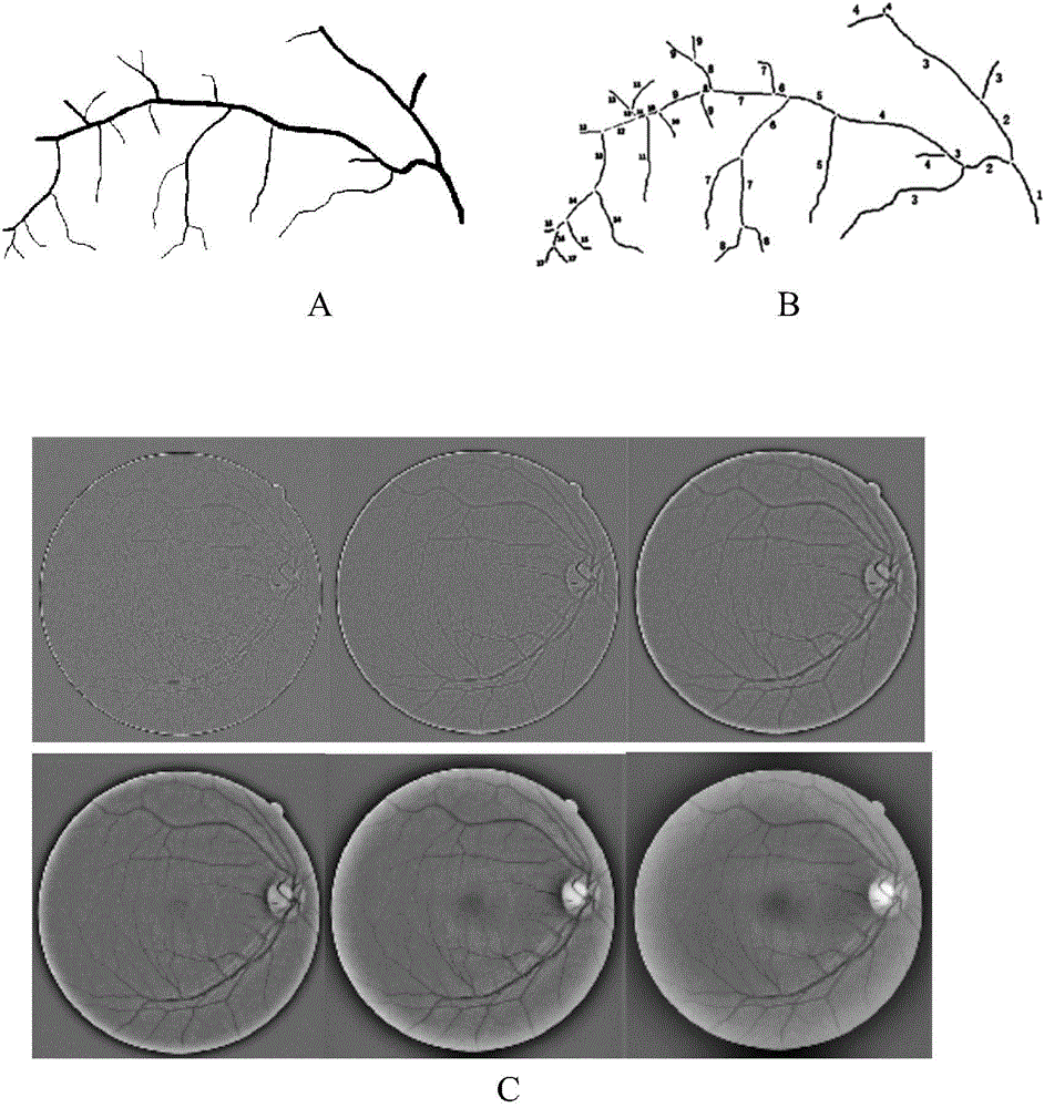
[0048] First, the green channel is extracted from the color RGB fundus image collected by the digital ophthalmoscope equipment (TOPCON, Japan), and then the grayscale conversion is performed using UWT transformation. After enhancement, the grayscale of pixels is extracted from the reconstructed image structure of the fourth layer. and local entropy features, using the FCM algorithm for automatic blood vessel segmentation, as figure 1 Segmentation results of the vessels shown.

[0049] In this experiment, the first non-subsampling discrete wavelet transform (UDWT) is used to decompose the two-dimensional image signal. The wavelet transform can obtain the time information of the signal by translating the basic wavelet (mother wavelet), and the frequency characteristics of the signal can be obtained by scaling the wavelet scale. Scaling and translation opera...

Embodiment 2

[0088] Embodiment 2: Using the method of the present invention to measure retinal vascular changes in early diabetic patients

[0089] Ten normal retinal images and 10 diabetic retinal images were used as the experimental objects, and the images were provided by the clinical ophthalmoscope workstation (Nantong University Affiliated Hospital). The images come from 20 patients with clinically diagnosed type 2 diabetes. The diagnostic criteria are "Diabetes Diagnostic Criteria Proposed by the American Diabetes Association in 1997." 2. Fasting blood sugar ≧7.0mmol / L. 3. Glucose tolerance test 2-hour blood glucose ≧ 11.1mmol / L. OGTT is still carried out in accordance with the requirements of the World Health Organization. Because there were 10 patients with hypertension, their fundus photographs were excluded. Finally, 10 images were included, including 6 males and 4 females, with an average age of 51.2 years. The fundus images of 10 cases were classified according to the severi...


PUM


Abstract

The invention belongs to the field of medical image processing and application, and provides a retinal vascular tortuosity calculation method based on an ophthalmoscope image and an application thereof. Firstly, a digital ophthalmoscope is used for obtaining an eye fundus image for screening the crowd, and a non-subsample discrete wavelet transform (UDWT) is then used for enhancing the image; secondly, local entropy texture of a retinal gray image is extracted, and a fuzzy C-mean clustering (FCM) method is used for segmenting a retinal vessel; and thirdly, the segmented vessel is skeletonized, topological levels of the skeleton are calculated, and a tortuosity calculation model in the invention is used for tortuosity calculation of the vascular skeleton. The method provided by the invention is easy to implement, accurate, reliable and convenient in clinical application.

Description

technical field [0001] The invention relates to the field of medical image processing and application, in particular to a method for calculating retinal blood vessel tortuosity based on ophthalmoscope images and its application. Background technique [0002] A typical retinal image includes the optic disc structure from which blood vessels and nerves enter the retina, the branches of blood vessels that emanate from the optic disc, and the macula in the center of the retina. Normal retinal vessels run straight or slightly curved, but dilate and tortuous in pathological conditions of high blood flow, angiogenesis, and vascular occlusion. Non-smooth vessel contouring, known as tortuosity, is usually the first change in vessel morphology. Clinically, tortuosity is an important index reflecting the severity of diabetic retinopathy. Curvature is an important attribute of morphological analysis. Tortuosity is usually described by the curvature change of blood vessels. Abnormal to...

Claims


Application Information

Patent Timeline
Patent Type & Authority Applications(China)
IPC IPC(8): G06T7/00
Inventor 吴辉群董建成耿兴云张晓峰
Owner NANTONG UNIVERSITY
Why Eureka
Try Eureka