HER2 nucleic acid aptamer and application thereof

A nucleic acid aptamer and nucleotide sequence technology, which can be used in pharmaceutical formulations, DNA/RNA fragments, biochemical equipment and methods, and can solve problems such as reducing the binding ability of nucleic acid aptamers and target molecules

Active Publication Date: 2015-11-25
THE INST OF BASIC MEDICAL SCI OF CHINESE ACAD OF MEDICAL SCI
View PDF1 Cites 10 Cited by
  • Summary
  • Abstract
  • Description
  • Claims
  • Application Information

AI Technical Summary

Problems solved by technology

Although the modification of the ribose 2' position can enhance the resistance of oligonucleotides to nucleases, it often reduces the binding ability of the nucleic acid aptamer to the target molecule

Method used


Image

  • HER2 nucleic acid aptamer and application thereof
  • HER2 nucleic acid aptamer and application thereof
  • HER2 nucleic acid aptamer and application thereof

Examples


Embodiment 1

[0058] The preparation of embodiment 1HB3-1, HB3

[0059] In the present invention, the core sequence of the HER2 nucleic acid aptamer is a single-stranded nucleotide with a length of 43 bases, and its nucleotide sequence is: 5'GTGTGTCTGCTATTTATTTTGCTTGATTATCTCTTAGGGATTT3'; the preferred HER2 nucleic acid aptamer is 59 bases The length of single-stranded nucleotides, the sequence is: 5'TGCCCGTGTCCCGAGGAGGTGCCCTATTTTGCTTGATTATCTCTAAGGGATTTGGGCGG-3'; more preferably, when synthesizing, all bases A in the above-mentioned sequence are modified with thio to obtain a thio HER Nucleic acid aptamers, respectively named (core sequence through thiol (HB3-1), preferred sequence through thiol (HB3)) The above sequences were all synthesized by Invitrogen. The structure of the HB3 aptamer is as follows figure 1 As shown, where A in the figure is the primary sequence, and B is the secondary structure.

[0060] Hereinafter, if there is no special statement, the terms "HB3" and "thio-HB3" bo...

Embodiment 2

[0061]Example 2 Ability of HB3-1 to bind to HER2 polypeptide

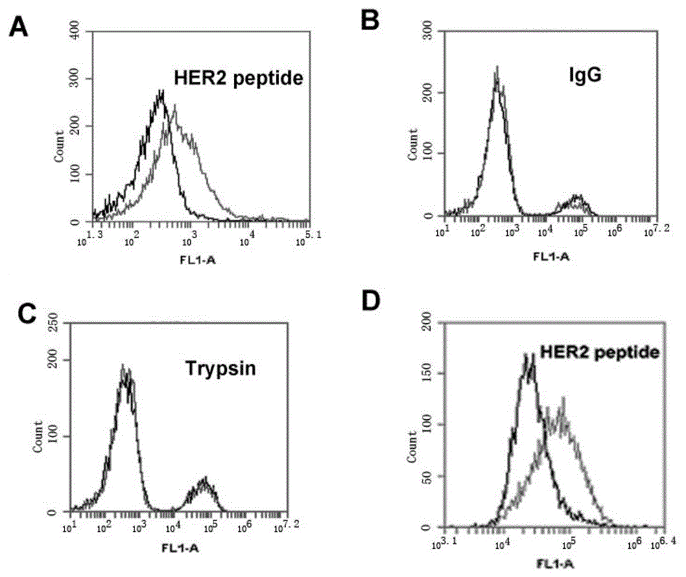
[0062] HB3-1 was modified with biotin (biotin) at the 3' end and FITC at the 5' end, and mixed with HER2 polypeptide, trypsin and The IgG-coated magnetic beads were reacted at 37°C for 30 minutes, washed twice with PBS, and analyzed by flow cytometry. The primary structure of the modified HB3-1 is as follows:

[0063] 5'FITC-GTGTGTCTGCTATTTATTTTGCTTGATTATCTCTTAGGGATTT-biotin-3'. Base A in the sequence is thio-modified.

[0064] Under the same conditions, we detected the binding of base A to the HER2 polypeptide without thio-modification.

[0065] The result is as figure 2 , wherein (A) the combination of HER2 polypeptide-coated magnetic beads and FITC-labeled HB3-1; (B) the combination of IgG antibody-coated magnetic beads and FITC-labeled HB3-1; (C) trypsin-coated Binding of magnetic beads and FITC-labeled HB3-1; (D) Binding of HER2 polypeptide-coated magnetic beads and FITC-labeled non-sulfo-modified HB3-1....

Embodiment 3

[0066] Example 3 Ability of HB3-1 to bind to HER2-positive tumor cells

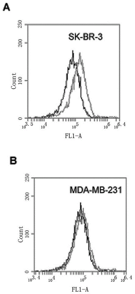
[0067] SK-BR-3 cells were cultured in modified RPMI1640 containing 15% FBS, MDA-MB-231 cells were cultured in DMEM high-glucose medium containing 10% FBS, 100 U / ml penicillin and 100 mg / ml streptomycin, All cells were stored at 37°C, 5% CO 2 Cultured in an incubator, the cells used in the following experiments were all in the logarithmic growth phase.

[0068] Scrape 5×10 separately 5 The SK-BR-3 and MDA-MB-231 cells were reacted with HB3-1 (modified biotin at the 3' end and fluorescein at the 5' end) in 200 μl binding buffer (PBS) at 37°C for 30 min, Wash twice with PBS and analyze by flow cytometry. see results image 3 , (A) is HER2 positive SK-BR-3 cells. (B) HER2-negative MDA-MB-231 cells. The black curve is the control fluorescence signal generated by random library single-stranded DNA, and the gray curve is the fluorescence signal generated by sulfo-HB3-1. The results in the accompanying dra...


PUM


Abstract

The invention relates to a nucleic acid aptamer specifically combined with HER2 polypeptide or HER2 positive tumor cells. Group modifying can be performed at two ends of the aptamer, group refers to one or multiple of amino, carboxyl, thiol, fluorescent molecule, cholesterol or polyethylene glycol, the base of the aptamer can be modified freely, and modifying refers to modifying one or multiple of sulfo, amino, fluoro, methoxyl or carboxyl. The invention further relates to application of the nucleic acid aptamer in preparing preparations used for detecting and / or treating HER2 positive breast cancer. The preparations can selectively combine with HER2 positive breast cancer cells while being weak in combination with HER2 negative breast cancer cells.

Description

technical field [0001] The invention belongs to the field of biotechnology, in particular, the invention relates to a nucleic acid aptamer capable of binding to human epidermal growth factor receptor 2 (HER2) molecules, in particular to a nuclease-resistant HER2 nucleic acid aptamer and the application of the nucleic acid aptamer in preparing preparations for detecting and / or treating HER2-positive breast cancer. Background technique [0002] Breast cancer is one of the common malignant tumors in women, about 20-30% of which are overexpressed human epidermal growth factor receptor 2 (HER2). Compared with other types of breast cancer, HER2-positive breast cancer has a higher degree of malignancy, faster progression, easier recurrence and distant metastasis, and lower sensitivity to chemotherapy drugs. In addition, more than 50% of HER2-positive breast cancers lack estrogen and progesterone receptors, so these patients are not sensitive to endocrine therapy. Since HR2-positi...

Claims


Application Information

Patent Timeline
Patent Type & Authority Applications(China)
IPC IPC(8): C12N15/115C12Q1/68A61K48/00A61P35/00
Inventor 杨先达胡燕
Owner THE INST OF BASIC MEDICAL SCI OF CHINESE ACAD OF MEDICAL SCI
Why Eureka
Try Eureka