

Method for establishing human knee joint three-dimensional simulation model in combination with CT (Computed Tomography) and MRI (Magnetic Resonance Imaging)
A two-dimensional image and three-dimensional simulation technology, applied in the field of medical imaging, can solve problems such as inability to understand the anatomical structure of the knee joint, and achieve clear anatomical relationships, convenient scanning, and clear images
- Summary
- Abstract
- Description
- Claims
- Application Information
AI Technical Summary
Problems solved by technology
Method used
Image

Examples
experiment example 1
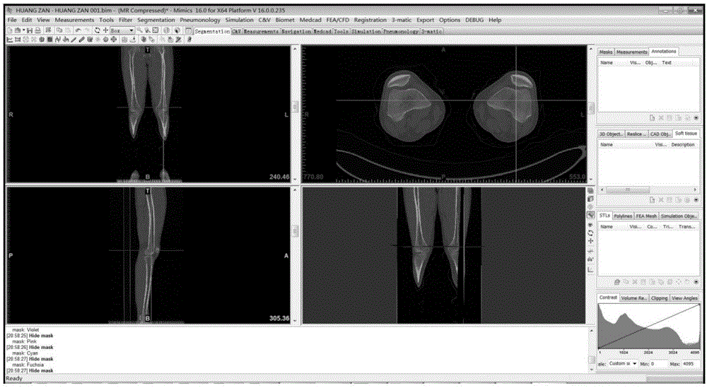
[0063] Establishment of 3D anatomical simulation model of human knee joint based on 2D images of CT and MRI
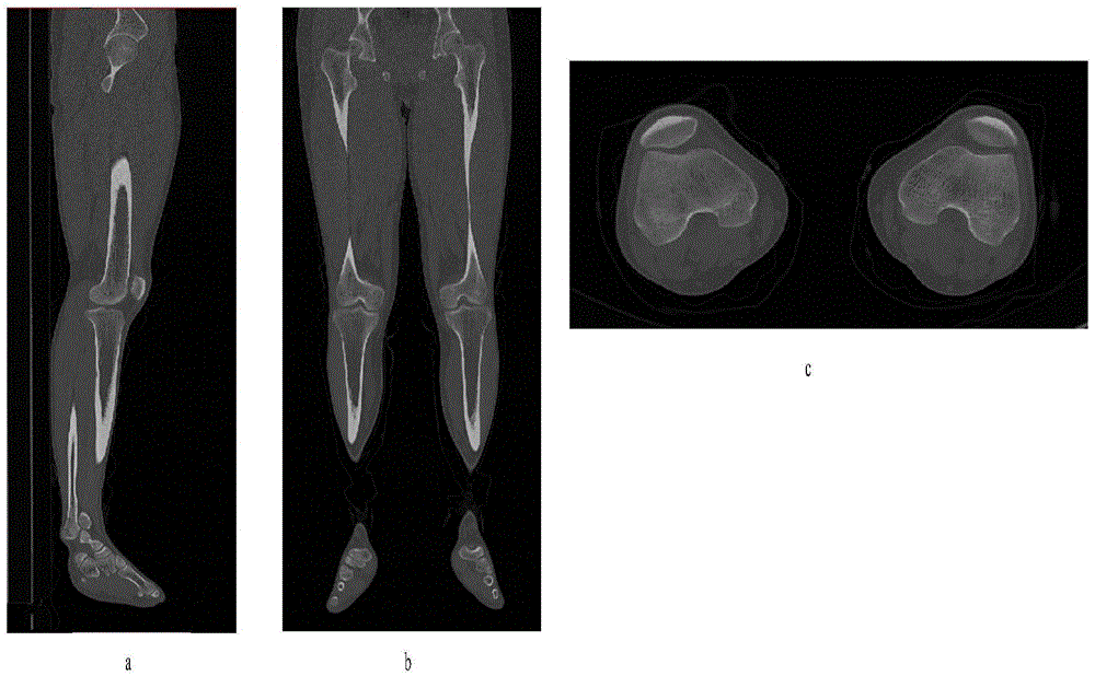
[0064] Step (1), CT image acquisition
[0065] SIEMENS 128-slice dual-source spiral CT was used to scan both lower extremities. Scanning position: the knee joint is naturally straightened and externally rotated at an angle of 10° to 15°. Scanning range: above to the middle pelvic plane, completely including the foot below. CT images are mainly used to observe bone tissue. Scanning parameters are set as follows: tube voltage of dual-source CT is 120kV and 70kV, tube current is 100mA and 60mA, slice thickness is 0.6mm, slice interval is 0.6mm, and pitch is 0.5.
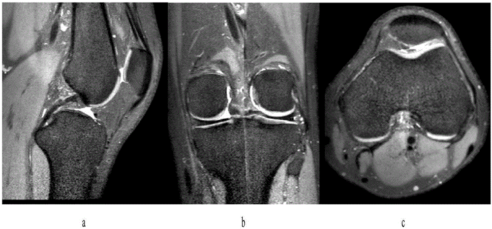
[0066] Step (2), MRI image acquisition
[0067] A GE1.5T superconducting magnetic resonance machine was used to scan both knees, and the magnetic resonance machine used the head coil as the receiving coil. The scanning position is that the knee joint is naturally straightened and externally rotated at an ang...
experiment example 2
[0085] Establishment of three-dimensional finite element model after artificial total knee arthroplasty
[0086] ① Prosthesis scanning and establishment of 3D digital model
[0087] After connecting the PFCSigma fixed platform total knee system prosthesis with the registration mark points, spray it in white and scan it with a 3D laser scanner, as shown in Figure 8 and Figure 9 shown. Import the scanned data into GeomagicStudio12 reverse engineering software, register the 3D point cloud model through the positioning points, and perform surface optimization. Firstly, the cavity on the model surface is filled based on the principle of curvature continuity, the non-characteristic indentation on the model surface is removed, the loose surface is smoothed, and the generation of non-characteristic high curvature and self-intersecting surfaces is prevented. Then use the contour line to describe the change of the surface curvature of the model, and use the parametric adjustment me...
experiment example 3
[0096] Dynamic finite element model analysis of artificial total knee arthroplasty
[0097] ① Coordinate calculation of the three-dimensional dynamic finite element model of the knee joint
[0098] According to the description of the flexion and extension, internal and external rotation, and adduction and abduction angles of the femur relative to the tibia, the three-dimensional dynamic model of the knee joint was established by using the definition method of the Cardan angle coordinate transformation. Define the coordinate system of the knee joint tibia in the straight position as the reference coordinate system 1, the knee joint femur coordinate system as the fixed coordinate system, and the other phase knee joint femoral coordinate system as the motion coordinate system 2, then the knee joint femur starts from 0° The Z-Y-X Euler angles of the extension coordinate system 1 transformed to other phase coordinate system 2 are φ, θ, ψ respectively, where φ represents the interna...
PUM
 Abstract
Description
Claims
Application Information