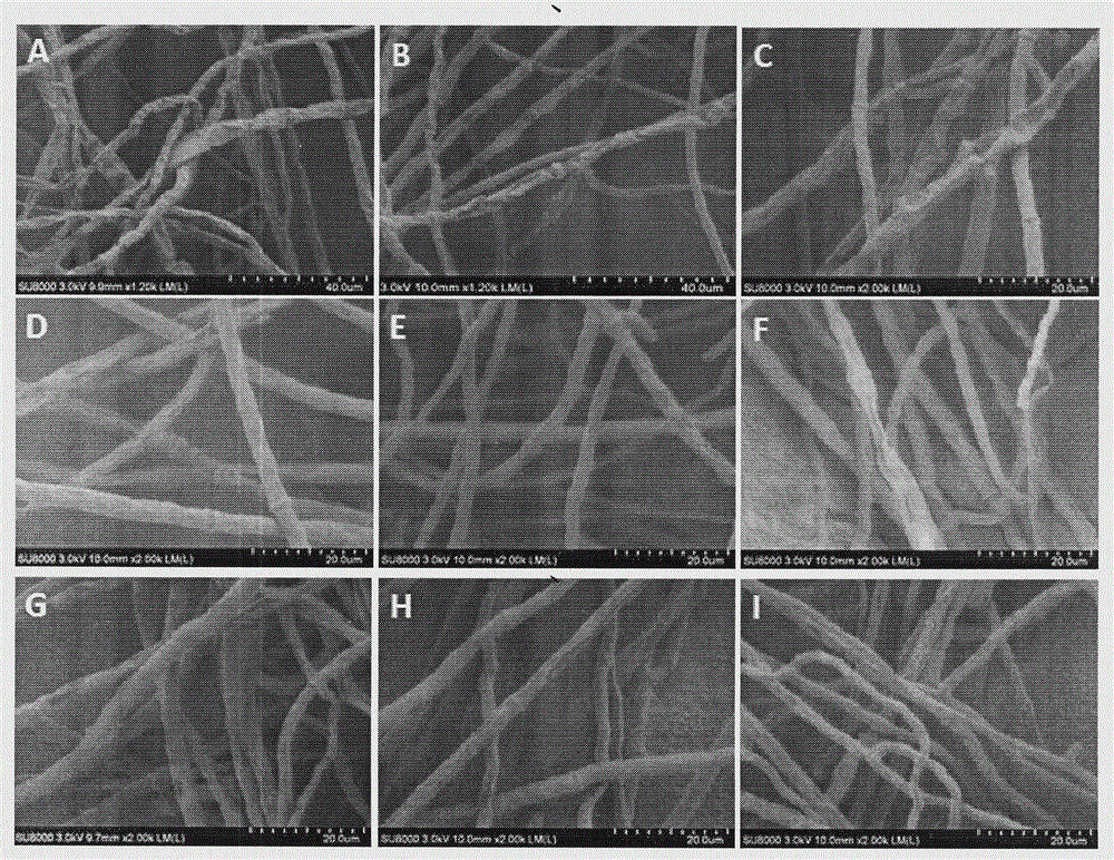
Method for preparing trichophyton sample for scanning electron microscope
A scanning electron microscope and Trichophyton technology, which is applied in the field of preparation of Trichophyton scanning electron microscope samples, can solve problems such as difficulty in obtaining results, shrinkage of bacteria, causing headache, inflammation of the respiratory tract, eye disease, bronchial disease, pneumonia and the like
- Summary
- Abstract
- Description
- Claims
- Application Information
AI Technical Summary
Problems solved by technology
Method used
Image
Examples
Embodiment 1
[0020] Embodiment 1: the preparation of scanning electron microscope sample of Trichophyton mentagrophytes
[0021] 1 Materials and reagents:
[0022] 1.1 Experimental material: Trichophytonmentagrophytes
[0023] 1.2 Reagents: peptone, glucose, agar, pH7.2 phosphate buffer solution (PBS), 25% glutaraldehyde, ethanol, acetone, tert-butanol
[0024] 1.3 Main experimental instruments: ultra-clean bench, incubator, pipette, refrigerator, autoclave, ultrasonic cleaner (AS3120A, 220V, 120W, Tianjin Autosines Instrument Co., Ltd.)
[0025] 1.4 Sample preparation
[0026] 1.4.1 Mycelia activation
[0027] SDA plate preparation: weigh 10g of peptone, 20g of glucose, and 18g of agar, dilute to 1L, sterilize at 121°C for 30min, pour the plate before the medium solidifies, and set aside;
[0028] Activation of strains: Inoculate the strains on a plate, culture in an incubator at 30°C for 5-7 days, and set aside.
[0029] 1.4.2 Sample preparation: inoculate the activated bacteria int...
PUM
 Abstract
Description
Claims
Application Information
 - R&D
- Intellectual Property
- Life Sciences
- Materials
- Tech Scout
- Unparalleled Data Quality
- Higher Quality Content
- 60% Fewer Hallucinations
Browse by: Latest US Patents, China's latest patents, Technical Efficacy Thesaurus, Application Domain, Technology Topic, Popular Technical Reports.
© 2025 PatSnap. All rights reserved.Legal|Privacy policy|Modern Slavery Act Transparency Statement|Sitemap|About US| Contact US: help@patsnap.com

