
Click chemistry-based tumor marker probe, preparation method and application

A click chemical reaction and tumor labeling technology, which is applied in the preparation method of peptides, chemical instruments and methods, applications, etc., can solve the problems of mutual interference and poor specificity, so as to avoid mutual interference, improve monitoring sensitivity, and improve bioavailability Effect

Inactive Publication Date: 2016-03-23
XIDIAN UNIV
View PDF1 Cites 13 Cited by
  • Summary
  • Abstract
  • Description
  • Claims
  • Application Information

AI Technical Summary

Problems solved by technology

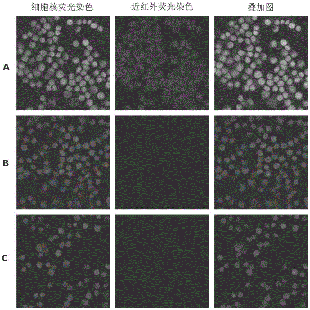
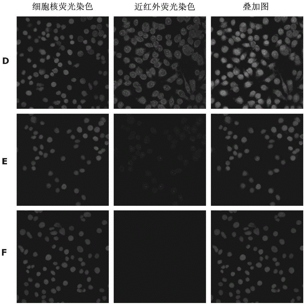
[0004] Aiming at the deficiencies of the prior art, the present invention aims to provide a tumor labeling probe based on click chemistry, a preparation method and its application. The tumor labeling probe includes click reaction modules with tumor-specific factors and fluorescent labels respectively. After the specific factors are pre-targeted to the tumor tissue, the fluorescent substances are labeled with the high selectivity, high efficiency and rapid response characteristics of click chemistry to avoid the mutual interference between the fluorescent substances and the specific factors, and solve the problem of existing tumor labeling probes or methods. The problem of poor specificity, and effectively reduce the metabolic clearance of probes or drugs by liver and kidney, thereby improving the bioavailability and monitoring sensitivity of probes

Method used


Image

  • Click chemistry-based tumor marker probe, preparation method and application
  • Click chemistry-based tumor marker probe, preparation method and application
  • Click chemistry-based tumor marker probe, preparation method and application

Examples


Embodiment 1

[0040] Preparation of tumor-specific factor click reaction module TCO-RGD:

[0041] Dissolve 0.64 mg of TCO-NHS in 128 μD DMSO in a 2 mL EP tube, add 70 μL of DMSO solution dissolved in 1.5 mg RGD peptide, then add 2 μLTEA, stir overnight at room temperature in the dark for 12 hours, and purify by separating out the product with diethyl ether and ethyl acetate The white powdery solid TCO-RGD was isolated, dried in vacuum and stored at low temperature. TCO-NHS is commercial grade, and can also be prepared by the following method: take TCO, add DIC and NHS, use DCM as the reaction solvent, stir at room temperature in the dark, and monitor by HPLC; after the reaction, use ether to separate out the product The solid was isolated by purification and dried in vacuo to obtain the product TCO-NHS.

Embodiment 2

[0043] Preparation of tumor-specific factor click reaction module TCO-GEBP11:

[0044] Dissolve 0.5 mg of TCO-NHS in 50 μD DMSO in a 2 mL EP tube, add 250 μL of DMSO solution dissolved with 2 mg of GEBP11 peptide, then add 4 μLTEA, stir at room temperature in the dark for overnight reaction (12 h), and use ether and ethyl acetate to separate out the product. The white powdery solid TCO-GEBP11 was obtained, which was vacuum-dried and stored at low temperature.

Embodiment 3

[0046] Preparation of fluorescently labeled click reaction module Tz-Cy5.5:

[0047] Take 1mg of Cy5.5 in a 2mL EP tube, add 7μLDIC, 4.6mg of NHS (the molar ratio of Cy5.5:DIC:NHS is 1:40:40), use 500μL of DMSO as the reaction solvent, and stir for 6h at room temperature in the dark. Monitored by reverse phase TLC. After the reaction, the product was purified and separated by diethyl ether and ethyl acetate to separate out the product, and dried in vacuum to obtain the product Cy5.5-NHS.

[0048] Dissolve 0.5 mg of Cy5.5-NHS in 300 μL DMSO in a 2mLEP tube, add 28 μL of DMSO solution with 0.28 mg Tz dissolved therein, then add 4 μLTEA (the molar feed ratio of Cy5.5-NHS:Tz is 1:3), and keep away from room temperature. Lightly stirred overnight (12h). After the reaction was completed, the mixture was purified and separated by washing with ether and ethyl acetate to obtain a blue solid, which was dried in vacuum and stored at low temperature.


PUM


Abstract

The invention discloses a click chemistry-based tumor marker probe, a preparation method and application. The tumor marker probe comprising a click reaction module with a tumor-specific factor and a click reaction module with a fluorescent marker is designed using reaction characteristics of click chemistry, such as high selectivity, high efficiency and high speed, and the two click reaction modules may be combined quickly through click reaction. The tumor marker probe is efficient in quickly combining to tumor tissues or cells, the life of the probe in viable tumor tissues and its detection time are effectively prolonged, the problem that an existing probe has poor specificity is effectively solved, and a new direction is provided for the in-vivo optical molecular imaging and live cell imaging of tumors as well as further targeted diagnosis and biological study of tumors.

Description

technical field [0001] The invention belongs to the field of molecular imaging applications, and relates to click chemistry-based tumor marker probes in near-infrared molecular imaging technology, a preparation method and applications thereof. Background technique [0002] Molecular imaging technology realizes real-time, non-invasive, and dynamic imaging of in vivo physiology and pathology at the level of cells and molecules, and can be used to study the occurrence, development, and metastasis of diseases or tumors in vivo. Among them, near-infrared fluorescent molecular imaging has non-radiation, sensitive High, light damage to biological tissue and other advantages, showing great potential in the field of tumor monitoring in vivo research. Near-infrared fluorescent probes realize the visual tracking and detection of target tissues, which is a prerequisite for the success of in vivo optical molecular imaging. Generally, fluorescent probes use diseased genes, molecules, tis...

Claims


Application Information

Patent Timeline
Patent Type & Authority Applications(China)
IPC IPC(8): C09K11/06C07K7/06C07K1/107C07D403/14A61B5/00
CPCC09K11/06A61B5/0071C07D403/14C07K1/1077C07K7/06C09K2211/1029C09K2211/1074
Inventor 张象涵王忠良赵娜王博夏玉琼田捷张瑞丽宁蓬勃胡波
Owner XIDIAN UNIV
Learn More