Molecular marker for diagnosing and treating hysteromyoma
A uterine fibroid, a pair of technology, applied in gene therapy, analytical materials, biological testing, etc., can solve the problems of patients feeling uncomfortable and poor sensitivity
- Summary
- Abstract
- Description
- Claims
- Application Information
AI Technical Summary
Problems solved by technology
Method used
Image
Examples
Embodiment 1
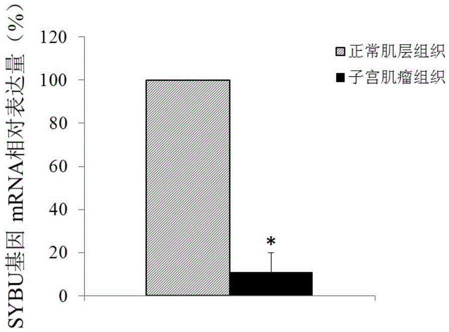
[0059] Example 1 Differences in the expression of SYBU gene in normal muscle layer tissue and uterine leiomyoma tissue
[0060] 1. Experimental materials:
[0061] Uterine fibroid tissue and adjacent normal muscle layer tissue were aseptically collected from patients undergoing total hysterectomy for uterine fibroids. The patient had not received hormone therapy within 3 months before operation, and all patients were pathologically diagnosed as uterine fibroids after operation. , the experimental materials were taken from 5 patients with uterine fibroids, and the patients were between 25 and 45 years old.
[0062] 2. Detection of differential expression of SYBU gene
[0063] 2.1 Extraction of tissue RNA
[0064] Use QIAGEN's RNApreppureTissueKit (Animal Tissue Total RNA Extraction Kit (DP431)) to extract according to the operating instructions.
[0065] 2.2 Quality analysis of RNA samples (NanoDrop1000 spectrophotometer)
[0066] NanoDrop1000 spectrophotometer detects RNA ...
Embodiment 2
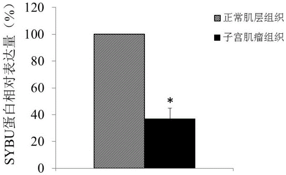
[0078] Example 2 Verification of Differentially Expressed Genes
[0079] 1. Experimental materials:
[0080] Uterine fibroid tissue and adjacent normal muscle layer tissue were aseptically collected from patients undergoing total hysterectomy for uterine fibroids. The patient had not received hormone therapy within 3 months before operation, and all patients were pathologically diagnosed as uterine fibroids after operation. , the experimental materials were taken from 50 patients with uterine fibroids, and the patients were between 25 and 45 years old.
[0081] 2. Detection of differential expression of SYBU gene
[0082] 2.1 Extraction of tissue RNA
[0083] Use QIAGEN's RNApreppureTissueKit (Animal Tissue Total RNA Extraction Kit (DP431)) to extract according to the operating instructions.
[0084] 2.2 Reverse transcription
[0085]1 μg of total RNA was reverse-transcribed to synthesize cDNA using reverse transcription buffer. Using a 25 μl reaction system, take 1 μg of...
Embodiment 3
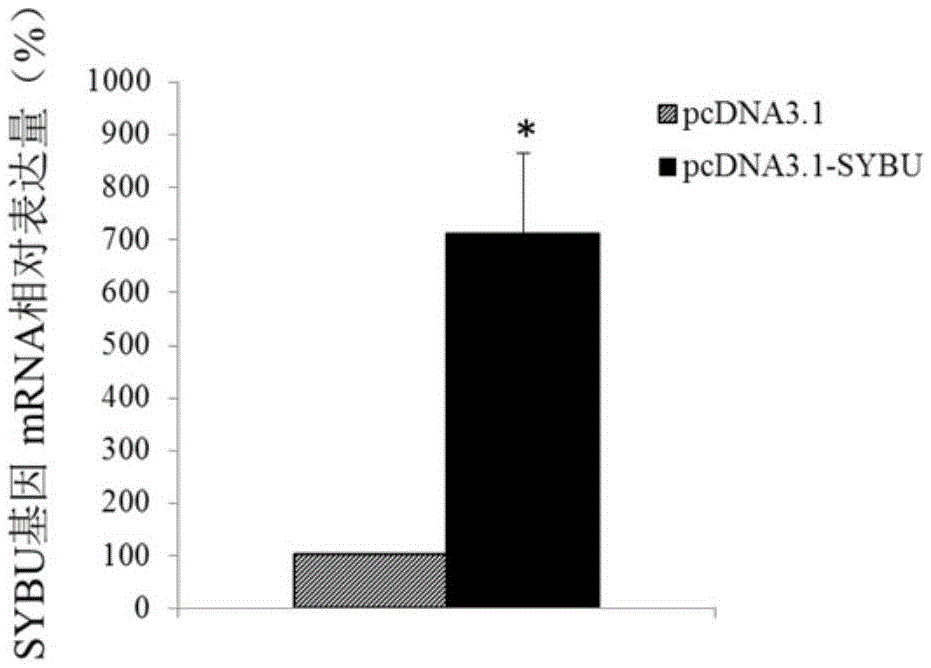
[0108] Example 3 SYBU gene expression plasmid construction
[0109] 1. Construction of SYBU gene expression vector
[0110] Amplification primers were designed according to the coding sequence of the SYBU gene (as shown in SEQ ID NO.1). Amplify the coding sequence of the full-length SYBU gene from the cDNA library of adult fetal brain (clontech company, article number: 638831), insert the above cDNA sequence into the eukaryotic cell expression vector pcDNA3.1, and connect the obtained recombinant vector pcDNA3.1 -SYBU was used in subsequent experiments.
[0111] 2. Culture and transfection of uterine leiomyoma cells
[0112] After surgical resection of uterine fibroids, 1 cm of uterine fibroid tissue was immediately taken under aseptic conditions, placed in 10 mL of 3% double-antibody (penicillin, streptomycin) PBS, and sent to the cell culture room as soon as possible in an airtight ice bath. Uterine leiomyoma tissue was rinsed with 3% double-antibody PBS solution, and tri...
PUM
 Abstract
Description
Claims
Application Information
 - R&D
- Intellectual Property
- Life Sciences
- Materials
- Tech Scout
- Unparalleled Data Quality
- Higher Quality Content
- 60% Fewer Hallucinations
Browse by: Latest US Patents, China's latest patents, Technical Efficacy Thesaurus, Application Domain, Technology Topic, Popular Technical Reports.
© 2025 PatSnap. All rights reserved.Legal|Privacy policy|Modern Slavery Act Transparency Statement|Sitemap|About US| Contact US: help@patsnap.com


