Rapid intraoperative pathological diagnosis system and method

A pathological diagnosis and rapid technology, applied in the field of medical information, can solve the problems of difficult control of production quality and slow production speed, and achieve the effect of complete preservation, avoiding time occupation, and rapid diagnosis

Active Publication Date: 2016-05-25
SHENZHEN INST OF ADVANCED TECH
View PDF10 Cites 7 Cited by
  • Summary
  • Abstract
  • Description
  • Claims
  • Application Information

AI Technical Summary

Problems solved by technology

[0004] In view of this, the embodiment of the present invention provides a system and method for intraoperative rapid pathological diagnosis to solve the problems of slow production speed and difficult control of production quality in the intraoperative pathological diagnosis method provided by the prior art

Method used


Image

  • Rapid intraoperative pathological diagnosis system and method
  • Rapid intraoperative pathological diagnosis system and method

Examples


Embodiment 1

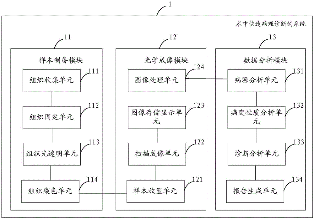
[0054] figure 1 A specific structural block diagram of the intraoperative rapid pathological diagnosis system provided by Embodiment 1 of the present invention is shown. For the convenience of description, only the parts related to the embodiment of the present invention are shown. The intraoperative rapid pathological diagnosis system 1 includes: a sample preparation module 11, an optical imaging module 12 and a data analysis module 13;

[0055] Wherein, the sample preparation module 11 includes:

[0056] Tissue collection unit 111, used to extract pathological sample tissue from the patient;

[0057] A tissue fixing unit 112, configured to fix the lesion sample tissue extracted by the tissue collection unit with chemical reagents;

[0058] A tissue light-transparency unit 113, configured to use a light-transparency agent to light-transparent the lesion sample tissue fixed by the tissue fixation unit;

[0059] The tissue staining unit 114 is configured to perform fluoresce...

Embodiment 2

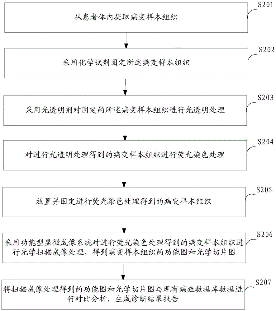
[0088] figure 2 It shows the implementation process of the method for intraoperative rapid pathological diagnosis provided by Embodiment 2 of the present invention, and is described in detail as follows:

[0089] In step S201, a lesion sample tissue is extracted from a patient.

[0090] In the embodiment of the present invention, the lesion sample tissue can be extracted from the patient's body to provide necessary materials for subsequent processing of the lesion sample tissue. Extraction operations include, but are not limited to, cutting, forceps, piercing, peeling, and the like.

[0091] In step S202, chemical reagents are used to fix the lesion sample tissue.

[0092] In the embodiment of the present invention, fixing the lesion sample tissue refers to rapidly coagulating the constituent substances in the lesion sample tissue and maintaining its structure in the living body. After fixing the lesion sample tissue, it can facilitate the subsequent light-transparent treat...


PUM


Abstract

The invention is suitable for the technical field of medical information and provides a rapid intraoperative pathological diagnosis system and method. The system comprises a sample preparation module, an optical imaging module and a data analysis module, wherein the sample preparation module extracts a diseased sample tissue from a patient body, fixes the diseased sample tissue by adopting a chemical reagent, then performs optical clearing treatment on the diseased sample tissue by adopting an optical clearing agent and finally performs fluorescent staining treatment on the diseased sample tissue; the optical imaging module first arranges and fixes the diseased sample tissue subjected to the fluorescent staining treatment, and then performs optical scanning imaging treatment on the diseased sample tissue subjected to the fluorescent staining treatment, by adopting a functional microimaging system, so as to obtain a functional diagram and an optical section diagram of the diseased sample tissue; and the data analysis module performs comparative analysis on the functional diagram and the optical section diagram as well as existing disease database data to generate a diagnosis result report. The system and the method are high in section-making speed and high in section-making quality.

Description

technical field [0001] The invention belongs to the technical field of medical information, and in particular relates to a system and method for intraoperative rapid pathological diagnosis. Background technique [0002] Pathological diagnosis is the most authoritative diagnosis in modern medicine at present. It refers to the technology of taking out diseased tissue from the patient's body for pathological examination in response to the needs of diagnosis and treatment. Through the analysis and identification of diseased tissue and cell morphology, combined with visual observation and clinical related data, various diseases can be diagnosed. [0003] Intraoperative pathological diagnosis, because of the short time required, can determine the benign and malignant lesions during the operation, and the diagnosis results can provide the basis for further clinical operation plans, and it is increasingly welcomed by the clinical operation departments of major hospitals at home and ...

Claims


Application Information

Patent Timeline
Patent Type & Authority Applications(China)
IPC IPC(8): G01N21/64G01N1/30
CPCG01N1/30G01N21/64
Inventor 郑炜陈廷爱张洋夏先园
Owner SHENZHEN INST OF ADVANCED TECH
Why Eureka
Try Eureka