
An In Vitro Method for the Label-Free Determination of the Cell Type of Cells

A cell-type technology, applied in the direction of individual particle analysis, using optical devices, instruments, etc., can solve problems such as limited reproducibility, low sample throughput, and continuous efforts to immobilize cells

Active Publication Date: 2019-04-02
SIEMENS HEALTHCARE GMBH
View PDF9 Cites 0 Cited by
  • Summary
  • Abstract
  • Description
  • Claims
  • Application Information

AI Technical Summary

Problems solved by technology

Drawbacks to this approach, however, are the low sample throughput compared to using hematology analyzers and the ongoing effort to immobilize and stain cells on microscope slides
Furthermore, these stains also have only limited reproducibility and exhibit a high dependence on humidity, stain life, temperature, etc.
The downside of pure microscopy is that it quantifies cell volume compared to flow cytometry used in hematology analyzers

Method used


Image

  • An In Vitro Method for the Label-Free Determination of the Cell Type of Cells
  • An In Vitro Method for the Label-Free Determination of the Cell Type of Cells
  • An In Vitro Method for the Label-Free Determination of the Cell Type of Cells

Examples


Embodiment Construction

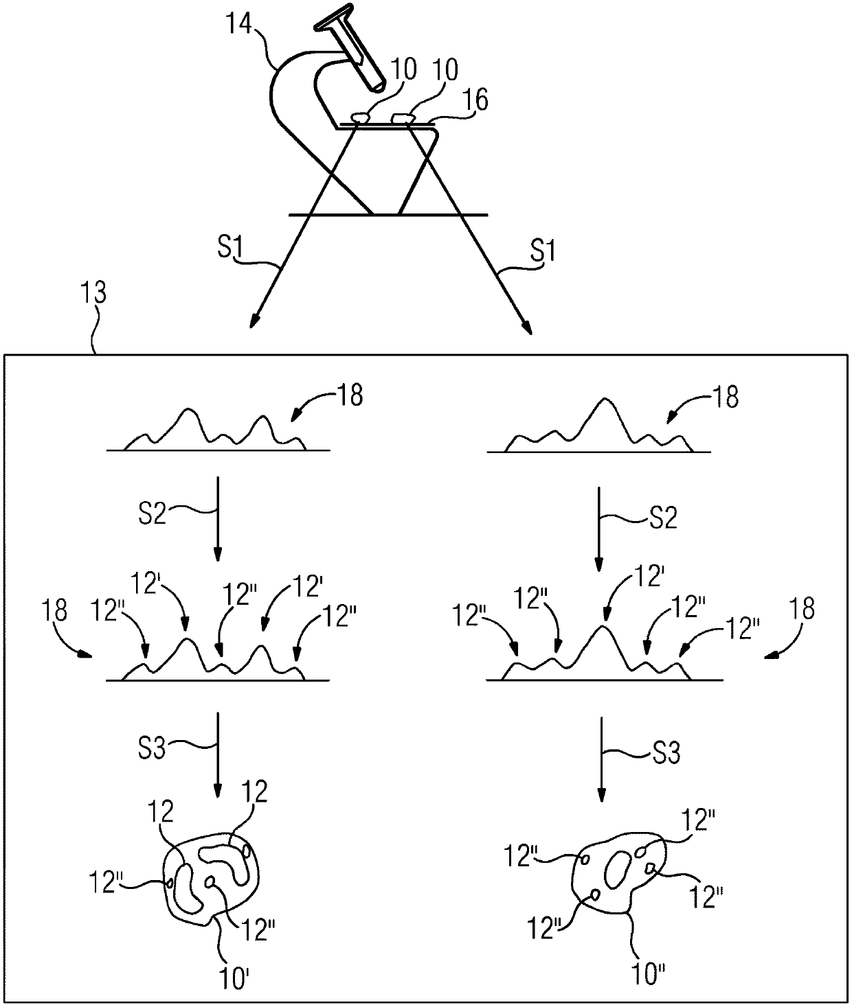
[0036] With the help of the inventive in vitro method, the cell type of cells in a biological sample can be determined in a marker-free manner. Label-free here means that the cells do not have to be labeled with eg fluorescent dyes or radioactive particles. A biological sample may eg comprise a sample of animal or plant cells, bacterial cells and / or protozoa. It is preferably a whole blood sample, comprising eg blood cells 10, eg leukocytes, eosinophils or basophils. For clarity, different exemplary cell types are in figure 1 with figure 2 , image 3 , Figures 4a and 4b are identified by the following reference numerals: eosinophil 10', basophil 10", neutrophil 10"', lymphocyte 10"" and monocyte"" '. However, for the sake of legibility, reference numeral 10 is used for each cell type in the following description of the exemplary embodiment.

[0037] exist figure 1 In the example of , for example, the five basic white blood cells that are distinguished in the case of a ...


PUM


Abstract

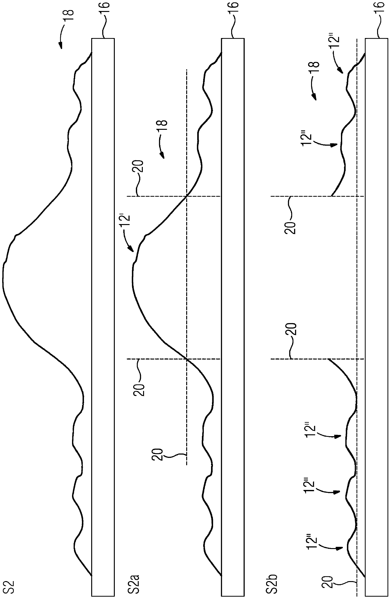
The invention relates to an in vitro method for the label-free determination of the cell type of cells (10) in a biological sample, wherein a microscopy device (14) detects (S1) the height profile (18) of the cells (10) relative to a support plate (16) . The method is characterized in that the cell analysis device (13) performs the following steps: determining the cell divisions (12, 12', 12") of the cell (10) on the basis of the detected height profile (18, S2). The predetermined quantitative cell characteristics are determined based on the cell divisions (12, 12', 12", S3), and the cell type of the cells (10) is determined based on the determined quantitative cell characteristics (S3). The invention also relates to a correspondingly designed cell analysis device (13) and a corresponding microscopy device (14).

Description

technical field [0001] The present invention relates to a method for the label-free determination of the cell type of cells using a microscope device. Background technique [0002] Identifying cells and assigning them cell types is very important in cytology. The blood cell components, ie erythrocytes, platelets and leukocytes, are determined and quantified eg in blood tests. Determination of the number of white blood cells (white blood cell count, WBC) should be able to distinguish the basic populations of white blood cells for a comprehensive diagnosis. Leukocytes are differentiated by granulosa cells (neutrophils, eosinophils, and basophils) and nongranulocytes (lymphocytes, monocytes). In addition to granularity cells are also concerned with fragmentation of the nucleus (without fragmentation: mononuclear cells, i.e. lymphocytes and monocytes; polymorphonuclear cells: eosinophils, basophils and neutrophils) and cell size varies. Staining is used to differentiate gran...

Claims


Application Information

Patent Timeline
Patent Type & Authority Patents(China)
IPC IPC(8): G01N15/10G01N15/14G01N15/00
CPCG01N2015/1454G01N15/10G01N2015/016G01N15/1433G01N15/01G01N2015/012G01B11/0608G01N2015/1493
Inventor O·海登O·施密特
Owner SIEMENS HEALTHCARE GMBH
Learn More