Method, oligonucleotide and kit for detecting was gene polymorphic mutation site
An oligonucleotide and mutation site technology, applied in the field of life science and biology, can solve the problems of easy misdiagnosis and complex clinical manifestations of WAS syndrome, and achieve the effect of high cost, high detection difficulty, and reduction of cost and difficulty.
- Summary
- Abstract
- Description
- Claims
- Application Information
AI Technical Summary
Problems solved by technology
Method used
Image

Examples
Embodiment 1
[0072] An oligonucleotide for detecting the polymorphic mutation site of the WAS gene, the oligonucleotide is designed for all exons of the WAS gene, including: amplification primers for detecting the polymorphic mutation site of the WAS gene, Its base sequence is:
[0073] WAS-1 / 2F: TGTAAAACGACGGCCAGTCAAAAGGTGGGTCTAAGCAGTC
[0074] WAS-1 / 2R: AACAGCTATGACCATGCGGGTTGAGAACTGGCTTG
[0075] WAS-3 / 4 / 5 / 6F: TGTAAAACGACGGCCAGTGAGCTGAAAATCTCCAAACCA
[0076] WAS-3 / 4 / 5 / 6R: AACAGCTATGACCATGTATCCATTCACCCACTTACGC
[0077] WAS-7F: TGTAAAACGACGGCCAGTTACCTCCATGACCATCCAACA
[0078] WAS-7R: AACAGCTATGACCATGCAGCCCTGCACCTACCTATC
[0079] WAS-8 / 9F: TGTAAAACGACGGCCAGTGAGGGCAAGAGGGTTTCACTA
[0080] WAS-8 / 9R: AACAGCTATGACCATGGCCTCAGTTTTGCTCATTTGT
[0081] WAS-10 / 11F: TGTAAAACGACGGCCAGTACCCCATTTTACAAATGAGCAAA
[0082] WAS-10 / 11R: AACAGCTATGACCATGGGTGACTGCTGGGATTGTTT
[0083] WAS-12F: TGTAAAACGACGGCCAGTTTCTTGTCCCAAATGGAAACTC
[0084] WAS-12R: AACAGCTATGACCATGGGCAGAAGGAAACAAAGAAATA.
[0085] In...
Embodiment 2
[0096] Operation process of blood / cell / tissue genomic DNA extraction kit (Tiangen Biology):
[0097](1) Extract tissue DNA from blood: 1) Extract 300 μl of blood and add 900 μl of erythrocyte lysate, mix by inverting, and place at room temperature for 5 minutes, during which time, invert and mix several times. Centrifuge at 12,000rpm for 1min, suck off the supernatant, leave the white blood cell pellet, add 200μl buffer GA, shake until thoroughly mixed. 2) Add 20 μl proteinase K solution and mix well. 3) Add 200 μl of buffer GB, mix thoroughly by inversion, place at 70°C for 10 minutes, the solution should become clear, and briefly centrifuge to remove water droplets on the inner wall of the tube cap. 4) Add 200 μl of absolute ethanol, vortex and mix well for 15 seconds. At this time, flocculent precipitates may appear. Briefly centrifuge to remove water droplets on the inner wall of the tube cap. 5) Add the solution and flocculent precipitate obtained in the previous step i...
Embodiment 3
[0123] Three cases of clinical blood samples (sample number 1-3) were taken according to the reagents and methods of Examples 1 and 2 to extract genome, prepare reagents, amplify and sequence. Each sample obtained its own DNA solution according to Example 2, and then added 1 μl of DNA solution to the PCR reaction solution of the detection system prepared according to Example 2, first amplified, and then electrophoresed the amplified product. Electrophoresis results such as figure 2 Shown, show that the primers of the present invention are paired to WAS-1 / 2F and WAS-1 / 2R, WAS-3 / 4 / 5 / 6F and WAS-3 / 4 / 5 / 6R, WAS-7F and WAS-7R, WAS- 8 / 9F and WAS-8 / 9R, WAS-10 / 11F and WAS-10 / 11R, WAS-12F and WAS-12R can effectively amplify blood samples with a single band.
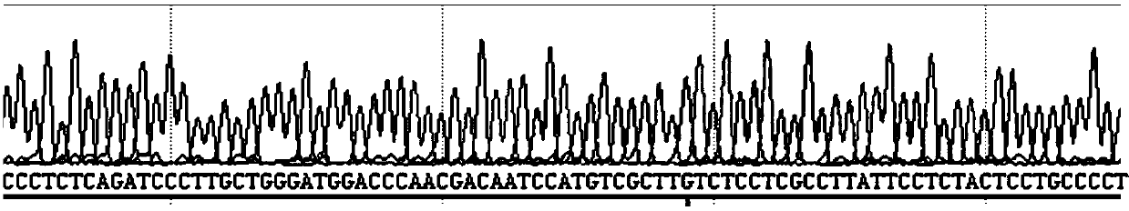
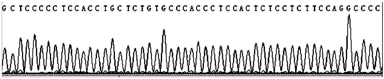
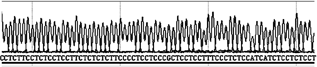
[0124] The test results of sample 1 are as follows: image 3 , 4 , 5, 6, 7, and 8 show:
[0125] image 3 It shows the wild-type sequencing screenshots of exons 1 and 2 of the WAS gene of sample 1, indicating that exons 1 and ...
PUM
 Abstract
Description
Claims
Application Information
 - R&D
- Intellectual Property
- Life Sciences
- Materials
- Tech Scout
- Unparalleled Data Quality
- Higher Quality Content
- 60% Fewer Hallucinations
Browse by: Latest US Patents, China's latest patents, Technical Efficacy Thesaurus, Application Domain, Technology Topic, Popular Technical Reports.
© 2025 PatSnap. All rights reserved.Legal|Privacy policy|Modern Slavery Act Transparency Statement|Sitemap|About US| Contact US: help@patsnap.com