CT image processing system and method for pneumoconiosis

A CT image and processing system technology, applied in the field of pneumoconiosis-oriented CT image processing system, can solve the problems of no pneumoconiosis cases, pneumoconiosis diagnosis and grading rely on the subjective judgment of doctors, image processing methods, etc.

Active Publication Date: 2016-07-13
SUZHOU INST OF BIOMEDICAL ENG & TECH CHINESE ACADEMY OF SCI
View PDF2 Cites 22 Cited by
  • Summary
  • Abstract
  • Description
  • Claims
  • Application Information

AI Technical Summary

Problems solved by technology

[0005] However, due to the various types and different classifications of pneumoconiosis, the appearance of pneumoconiosis on CT images is diverse, making the diagnosis and classification of pneumoconiosis basically rely on the subjective judgment of doctors
At present, there are many image processing methods such as pulmonary nodule segmentation for CT images, but there is no image processing method specifically for pneumoconiosis cases.

Method used


Image

  • CT image processing system and method for pneumoconiosis
  • CT image processing system and method for pneumoconiosis
  • CT image processing system and method for pneumoconiosis

Examples


Embodiment 1

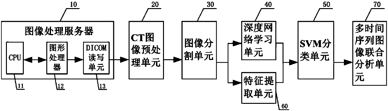
[0063] Such as figure 1 As shown, the present invention provides a pneumoconiosis-oriented CT (Computed Tomography, computerized tomography) image processing system, including an image processing server 10, which includes a CPU 11, a graphics processor 12, and a DICOM (Digital Imaging and Communications in Medicine, Medical digital imaging and communication) read-write unit 13; PACS (PictureArchivingandCommunicationSystems, audio-visual archiving and finishing system) system is installed in the graphics processor 12, and the data transmission between CPU11 and graphics processor 12 is through PCI-E (PCIExpress, bus interface ) is completed; DICOM read-write unit 13 reads and analyzes the CT image of pneumoconiosis from the PACS system of graphic processor 12; Preprocessing of correction, image denoising and artifact removal; image segmentation unit 30, which performs lung parenchyma segmentation, lung nodule segmentation and lung nodule false positive target removal on the pre...

Embodiment 2

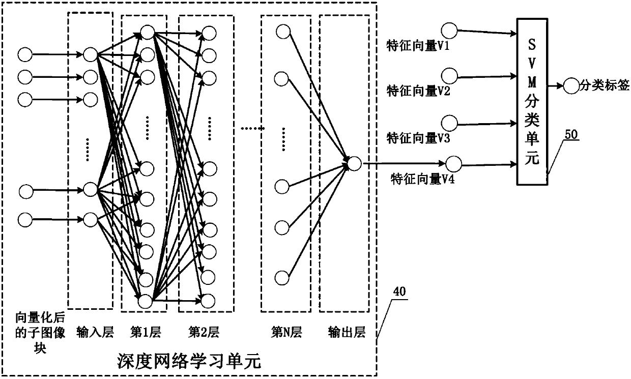
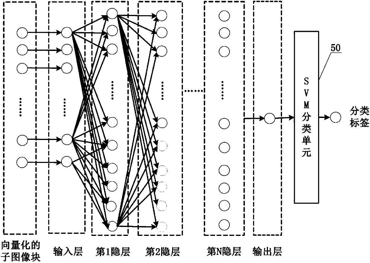
[0069] On the basis of Embodiment 1, this embodiment provides a pneumoconiosis-oriented CT image processing method, including the following steps: DICOM read-write unit 13 reads the CT image from the graphics processor 12 and parses it into a three-dimensional volume image; The CT image preprocessing unit 20 performs preprocessing of grayscale unevenness correction, image denoising and artifact removal on the three-dimensional volume image; the image segmentation unit 30 performs lung parenchyma segmentation, pulmonary nodule segmentation and pulmonary nodule segmentation on the preprocessed CT image. Nodule false positive target removal; the deep network learning unit 40 extracts the high-dimensional features in the sub-image block where the pulmonary nodule area segmented by the image segmentation unit is located; the SVM classification unit 50 receives the high-dimensional features for classification.

[0070] The pneumoconiosis-oriented CT image processing method provided b...

Embodiment 3

[0091] In the pneumoconiosis-oriented CT image processing method, the learning process of the deep network learning unit 40 needs to calculate the data of a large number of nodes in multiple levels, resulting in a very slow learning speed of the deep network learning unit 40 . On the basis of Embodiment 2, the pneumoconiosis-oriented CT image processing method provided in this embodiment also includes outputting the feature vector V4 through a parallel acceleration method, such as Figure 4 As shown, it specifically includes the following steps:

[0092] Carry out data segmentation, divide the same CT image into I sub-image blocks according to the total number of pulmonary nodules in the CT image, and extract the sub-image block G that can cover the largest pulmonary nodule area i , (i=1,2,...,I), each sub-image block has at most N 3 threads; each sub-image block has an exclusive video memory space, and a deep network model is trained correspondingly in each sub-image block; ...


PUM


Abstract

The invention discloses a CT image processing system for pneumoconiosis, and the system comprises an image processing server which comprises a CPU, a graphic processor and a DICOM reading-writing unit connected to the graphic processor, wherein the DICOM reading-writing unit reads a CT image from the graphic processor and analyzes the CT image; a CT image preprocessing unit which carries out the gray scale inhomogeneity correction, denoising and artifact removing of the CT image analyzed by the DICOM reading-writing unit; an image segmenting unit which carries out the pulmonary parenchyma segmentation, pulmonary nodule segmentation and pulmonary nodule false positive target segmentation of the CT image after preprocessing; a depth network learning unit which extracts the high-dimensional features of a sub-image block where a pulmonary nodule region is located after the segmentation; and an SVM classification unit which receives the high-dimensional features for classification. The system is high in classification precision of the data of the CT image, and is stable in robustness.

Description

technical field [0001] The invention relates to the technical field of graphic processing, and more specifically, the invention relates to a pneumoconiosis-oriented CT image processing system and a method thereof. Background technique [0002] A large amount of dust accumulates in the lung tissue, causing pathological changes of lung tissue fibrosis and progressive damage to the lung tissue. According to data from the Ministry of Health, by the end of 2009, a total of 722,730 cases of occupational diseases had been reported nationwide, including 653,000 cases of pneumoconiosis, accounting for more than 90% of the total number of occupational diseases. Statistics in 2010 showed that there were 23,812 new cases of pneumoconiosis in my country, and 190,000 people were identified as "observed objects" of pneumoconiosis, which became the main component of new pneumoconiosis cases. Cases of pneumoconiosis in my country are on the rise, causing annual direct economic losses of mor...

Claims


Application Information

Patent Timeline
Patent Type & Authority Applications(China)
IPC IPC(8): G06K9/46G06K9/62
CPCG06T5/00G06T5/002G06T5/40G06T15/005G06T2200/04G06T2207/10081G06T2207/30064G06V10/40G06V2201/031G06F18/241
Inventor 佟宝同周志勇耿辰胡冀苏刘燕戴亚康
Owner SUZHOU INST OF BIOMEDICAL ENG & TECH CHINESE ACADEMY OF SCI
Why Eureka
Try Eureka