A low-temperature endoscope blood vessel collection device and method
A collection device and endoscope technology, applied in the medical field, can solve the problems of great pain, intense pain, and long recovery time, and achieve the effects of reducing the chance of infection, shortening the recovery time, and reducing pain
- Summary
- Abstract
- Description
- Claims
- Application Information
AI Technical Summary
Problems solved by technology
Method used
Image

Examples
Embodiment 1
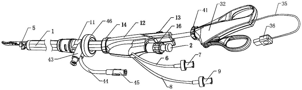
[0045] Embodiment 1: A low-temperature endoscopic blood vessel collection device is composed of a rigid tube endoscope 2, a short-mouth trocar 11, a free sleeve, a collection sleeve, bipolar scissors, and low-temperature electrocoagulation scissors;
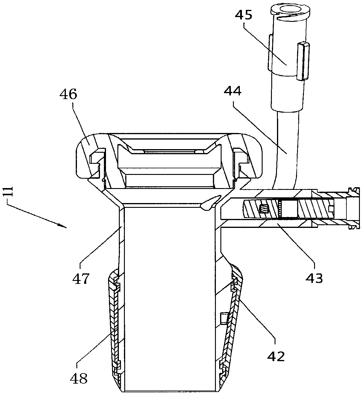
[0046] Such as figure 1 As shown, the connection port trocar 11 of the collection cannula 1, one end of the cannula 1 is connected with the connecting mechanism 14, the connecting mechanism 14 is fixed on the handle 12, and the handle 12 is connected with the sliding button 13; 2 is locked on the handle 12, and the bipolar scissors are inserted into the through holes 10 of the scissors.
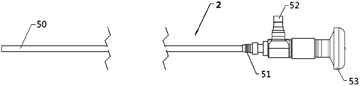
[0047] exist figure 2 In the schematic diagram of the structure of the rigid tube endoscope 2 shown: the rigid tube endoscope 2 is made of a steel pipe 50, the tip of the steel tube 50 is a lens for collecting images, and the end has a thread 51 for connecting with the collection sleeve and the free sleeve. fixed. The endoscope is provided w...
Embodiment 2
[0053] Embodiment 2: The rigid tube endoscope has an effective length of 20-45 cm, a diameter of 3-8 mm, an optical working distance of 20 mm, an effective depth of field of 3-100 mm, a viewing angle of 0° and a field of view of 75°.
[0054] The length of the annular air bag of the short-mouth trocar is 1-3cm, and the diameter after inflation is 2-5cm; the main body seal has two sizes, one with a diameter of 6-15mm for matching with the collection cannula; the other with a diameter of 3 -10mm is used to cooperate with the free sleeve 20.
[0055] The effective length of the free casing is 25-40cm; the outer diameter of the free casing is 5-15mm; the outer diameter of the tapered head is 9-18mm.
[0056] The effective length of the collection sleeve is 20-45cm, and the outer diameter is 9-20mm; the outer surface of the sleeve is a prismatic surface; the diameters of the scissors through hole and the endoscope through hole are respectively 4-9mm; the length of the handle is 8-1...
Embodiment 3
[0059] Embodiment 3: A method for collecting blood vessels through a low-temperature endoscope, comprising the following steps;
[0060] First insert the endoscope 2 into the free sleeve 20, and install the free head 21;
[0061] Put the short-mouthed trocar 11 on the cannula 20, insert the free head 21 into the surgical site through the short-mouthed trocar 11, and push it toward the target;
[0062] After the free head 21 reaches the operation position, take out the free sleeve 20, and pull out the endoscope 2 therefrom;
[0063] Insert the endoscope 2 into the endoscope through hole 3 of the collection cannula, and insert low-temperature electrocoagulation scissors or bipolar scissors into the scissors through hole 10. Insert the collection cannula through the short-mouth trocar 11 into the surgical site;
[0064] Under the monitoring of the endoscope 2, push the c-ring 5 to the target blood vessel. If the lens is blurred by blood or fat, connect the cleaning solution inje...
PUM
 Abstract
Description
Claims
Application Information
 - R&D
- Intellectual Property
- Life Sciences
- Materials
- Tech Scout
- Unparalleled Data Quality
- Higher Quality Content
- 60% Fewer Hallucinations
Browse by: Latest US Patents, China's latest patents, Technical Efficacy Thesaurus, Application Domain, Technology Topic, Popular Technical Reports.
© 2025 PatSnap. All rights reserved.Legal|Privacy policy|Modern Slavery Act Transparency Statement|Sitemap|About US| Contact US: help@patsnap.com