Method for separating and extracting exosomes in lamination centrifugal-filtration mode

A technology of centrifugal filtration and exosomes, which is applied in the field of molecular biology and clinical testing, can solve the problems of limited number of parallel processing samples, lengthy operation process, unfavorable clinical application, etc., and achieve the effect of low cost of consumables and shortened operation time

Active Publication Date: 2016-11-16
东莞微量精准检测研究院有限公司
View PDF7 Cites 21 Cited by
  • Summary
  • Abstract
  • Description
  • Claims
  • Application Information

AI Technical Summary

Problems solved by technology

[0006] 1) The operation takes a long time: the whole process from the processing of biological fluid to the collection of exosomes takes about 8-24 hours;
[0007] 2) Low separation efficiency: the academic community has proven that the recovery rate of exosomes by this method is only about 10% [5] , a large sample size is required to obtain sufficient samples for analysis;
[0008] 3) Specific instruments are required: the ultracentrifuge used for extraction is not a conventional laboratory equipment;
[0009] 4) The number of samples processed in parallel is limited: according to different centrifuge rotors, the number of samples that can be operated in parallel at the same time is generally not more than 6
[0012] 1) High experimental cost: Taking mainstream commercial reagents, such as System Biosciences (SBI)’s ExoQuick-TC as an example, the reagent consumption for each extraction exceeds RMB 180, which is not conducive to large-scale promotion and clinical application;
[0013] 2) Subsequent processing is limited: since the extraction process requires adding additional components to the sample, the collected exosomes cannot be directly used for cell culture or reinfusion;
[0014] 3) Not suitable for large-volume samples: due to its principle and high cost, this method is not suitable for the processing of large-volume samples
[0017] 1) High experimental cost: magnetic beads require special processing capabilities, and the simultaneous extraction of the required antibodies and cross-linking of antibodies and magnetic beads further increases the implementation cost of this method;
[0018] 2) Only a specific subpopulation of exosomes in the sample can be extracted: Since this method is based on antigen-antibody reactions, only exosome subpopulations with a known membrane surface antigen can be specifically extracted from the sample;
[0019] 3) Not suitable for large-volume samples: due to its principle and high cost, this method is not suitable for the processing of large-volume samples
[0020] 4. Some studies have also used ultrafiltration for exosome enrichment [12,13] , but the reported method requires ultracentrifugation to further precipitate exosomes, the operation process is lengthy, and it is not suitable for clinical use
And there is no method that can be applied to the ultrafiltration of serum / plasma to extract exosomes
[0021] To sum up, there is currently no exosome extraction method that is cheap, convenient to operate, short in time, and can perform a large number of sample operations in parallel to meet the needs of large-scale clinical application.

Method used


Image

  • Method for separating and extracting exosomes in lamination centrifugal-filtration mode
  • Method for separating and extracting exosomes in lamination centrifugal-filtration mode
  • Method for separating and extracting exosomes in lamination centrifugal-filtration mode

Examples


Embodiment 1

[0089] Example 1 A stacked centrifugal filter device for separating and extracting exosomes

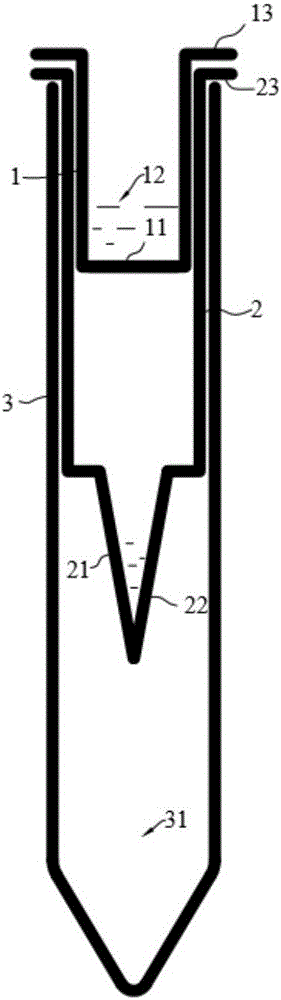
[0090] The invention provides a stacked centrifugal filter device for separating and extracting exosomes (see figure 1 ), comprising a filter tube 1, an ultrafiltration tube 2 set on the outside of the filter tube 1 and a waste liquid collection tube 3 set on the outside of the ultrafiltration tube 2, the bottom of the filter tube 1 is provided with a filter membrane 11, and the inside of the filter tube 1 A primary filter lumen 12 is formed, an ultrafiltration membrane 21 is provided at the bottom of the ultrafiltration tube 2, a secondary filter lumen 22 is formed inside the ultrafiltration tube 2 below the filter tube 1, and the waste liquid collection tube 3 is inside the ultrafiltration tube The bottom of the filter tube 2 forms a waste liquid collection lumen 31, the top nozzle of the filter tube 1 is provided with a primary support edge 13, the top nozzle of the ultrafiltration...

Embodiment 2

[0099] Example 2 A kit for separating and extracting exosomes

[0100] The kit includes the following components:

[0101] 1) Laminated centrifugal filter device, as described in Example 1.

[0102] 2) Incubation buffer, including the following components: DPBS, 60-240 mM Tris·HCl buffer, 60-180 mM EDTA, pH 7-7.5.

[0103] 3) Proteinase K stock solution, the concentration is 5-40mg / mL.

[0104] 4) DPBS: KCl 200mg / L; KH 2 PO 4 200mg / L; NaCl 8g / L; NaCl 2 HPO 4 ·7H 2 O 2.16g / L.

Embodiment 3

[0105] Example 3 A method for separating and extracting exosomes

[0106] Exosomes were isolated and extracted using the kit of Example 2.

[0107] Include the following steps:

[0108] 1) Add 0.5 times the volume of incubation buffer to the sample to be tested, and add 0.05 times the volume of proteinase K stock solution, mix well, and incubate at room temperature for 8-15 minutes;

[0109] 2) Transfer the sample processed in step 1) to a stacked centrifugal filter device (see figure 1 ) filter tube, centrifuge, mix, and collect the retentate in the ultrafiltration tube.

[0110] Preferably, if you need to improve the purity of exosomes, add DBPS to the ultrafiltration tube, centrifuge at 3,000×g for 10 minutes to remove some soluble proteins, discard the filtrate and repeat this step once.

[0111] Preferably, mixing and collecting the retentate is the exosome solution, which can be processed later.

[0112] The method of the present invention is compared with the prior ...


PUM

PropertyMeasurementUnit
pore sizeaaaaaaaaaa
diameteraaaaaaaaaa
diameteraaaaaaaaaa

Abstract

The invention discloses a method for separating and extracting exosomes in a lamination centrifugal-filtration mode. The method is used for the technical field of molecular biology and clinical examinations. A device comprises a lamination centrifugal-filtration device for separating and extracting the exosomes and an exosome separating and extracting kit composed of an incubating buffering solution and protease K. To-be-measured samples are incubated through the incubating buffering solution and the proper amount of protease K in the kit at the room temperature, then centrifugation is carried out through the lamination centrifugal-filtration device in a matched centrifugal machine, the product is mixed to be even, trapped liquid in an ultrafiltration pipe is collected, and the exosomes can be obtained. According to the method, in addition to the centrifugal machine, large experimental devices are not required, cost is low, operation is convenient and fast, time is short, a large amount of sample operation can be parallelly carried out, and the obtained high-purity exosomes can meet the requirements of clinical large-scale application and popularization.

Description

technical field [0001] The invention belongs to the field of molecular biology and clinical examination, and in particular relates to a method for separating and extracting exosomes by lamination centrifugal filtration. Background technique [0002] Exosomes (exosomes) are nanoscale membranous vesicles released into the extracellular environment after the fusion of multivesicular bodies (MVBs) and cell membranes of eukaryotic cells, with a lipid bilayer membrane. Structure, about 30–150nm in size, widely distributed in body fluids; can be secreted by various types of cells, which contain different types of proteins, lipids, mRNAs, microRNAs and signal molecules and other biologically active substances; strong Targeting, easy to fuse with the cell membrane of target cells, selectively deliver biologically active substances to target cells, transmit information between different cells, regulate intercellular signal transduction, and exert various biological functions. [0003...

Claims


Application Information

Patent Timeline
Patent Type & Authority Applications(China)
IPC IPC(8): G01N1/34
CPCG01N1/34B01L3/5021B01L2200/0684B01L2300/048B01L2300/0681B01D63/06B01D63/087B01D61/18B01D63/16G01N1/4077G01N2001/4088C12Q1/00B01D61/146B01D61/145B01D69/02B01D2325/02B01D2325/34
Inventor 王通崔毅峙罗彦彰郭嘉慧
Owner 东莞微量精准检测研究院有限公司
Why Eureka
Try Eureka