
Method of analyzing protein secondary structure in peony on the basis of infrared spectroscopy and Gauss multi-peak fitting

A technology of secondary structure and analysis method, applied in the field of spectral analysis, can solve the problems of long period, narrow data coverage, high cost, and achieve the effects of high accuracy, shortened experimental period, and strong data convincing.

Inactive Publication Date: 2016-12-07
INST OF FORESTRY & FRUIT TREE WUHAN ACADEMY OF AGRI SCI & TECH
View PDF4 Cites 2 Cited by
  • Summary
  • Abstract
  • Description
  • Claims
  • Application Information

AI Technical Summary

Problems solved by technology

[0005] In view of the deficiencies in the prior art, the purpose of the present invention is to provide a simple and effective method for detecting the secondary structure of all proteins in tree peony, so as to fill the gap in the study of protein secondary structure at the plant level and solve the problem of high cost and cycle time of the prior art. Difficulties plaguing researchers such as length and narrow data coverage

Method used


Image

  • Method of analyzing protein secondary structure in peony on the basis of infrared spectroscopy and Gauss multi-peak fitting
  • Method of analyzing protein secondary structure in peony on the basis of infrared spectroscopy and Gauss multi-peak fitting
  • Method of analyzing protein secondary structure in peony on the basis of infrared spectroscopy and Gauss multi-peak fitting

Examples


Embodiment 1

[0022] Example 1: Identification and analysis of protein secondary structure in peony

[0023] The dried peony root sample was ground into powder, and the potassium bromide tablet method was used for FTIR scanning. Perform Gaussian single-peak fitting on the original spectrum, and then perform Gaussian multi-peak fitting on the fitted single peak according to the peak positions of the characteristic peaks of different protein secondary structures, and calculate the percentage of various protein secondary structures in the sample according to the peak area.

[0024] The specific operation method is as follows:

[0025] (1) Take 1-2 mg of dried peony root and 200 mg of dried KBr and grind it into a uniform fine powder (about 2 μm in diameter) to avoid the influence of scattered light. Put the bottom mold into the mold cavity (note that the polished side is upward), add a small amount of sample to the mold core, scrape the sample in the mold with a medicine spoon, and make the c...

Embodiment 2

[0029] Example 2: Protein secondary structure analysis in peony root

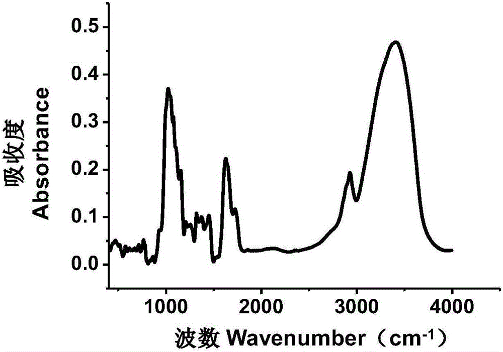
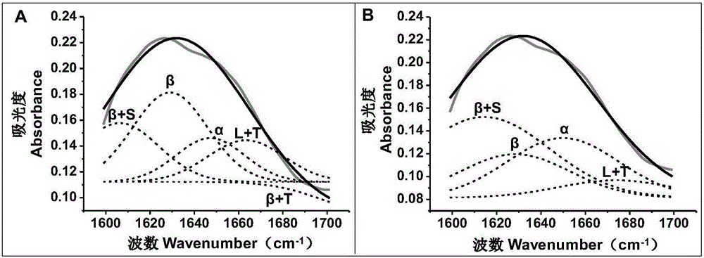
[0030] The operation steps described in Example 1 were selected to analyze the secondary structure of the protein in the peony root. The FTIR curve of peony root is as follows figure 1 As shown, it shows that there is a peak of amino I in peony root, that is, at 1600-1700cm -1 There is an infrared absorption peak. For 1600-1700cm -1 spectrogram ( figure 2 The gray solid line in Figure A and Figure B) is used for Gaussian unimodal fitting, and the results are as follows figure 2 As shown by the solid black lines in Figures A and B, the fitting equation is:

[0031] y = y 0 + ( A w × π 2 ) - ...

Embodiment 3

[0036] Example 3: Analysis of protein secondary structure in peony branches

[0037] Dry peony branch is ground into powder, and specific operation method is according to the operation method of embodiment 1. FTIR results of peony branches show at 1600-1700cm -1 There is an infrared absorption peak at , and its Gaussian single-peak fitting equation is:

[0038] y = y 0 + ( A w × π 2 ) - 2 × ( x - x c w ...


PUM

PropertyMeasurementUnit
particle diameteraaaaaaaaaa
diameteraaaaaaaaaa

Abstract

The invention discloses a method of analyzing a protein secondary structure in peony on the basis of infrared spectroscopy and Gauss multi-peak fitting. The method includes the steps of: grinding a dry peony sample into powder and performing FTIR scanning through a potassium bromide tabletting method; performing Gauss single-peak fitting and performing Gauss multi-peak fitting to the fit single peaks according to characteristic peak positions of different protein secondary structures; and calculating the percentage ratios of various protein secondary structures in the sample according the peak area. The method has the following advantages: 1) the method allows the analysis on the protein secondary structure directly on the level of the plant without extraction and purification of proteins, so that experimental cost is greatly reduced and experimental period and labor and material resources are reduced; and 2) data collection and analysis in the method are based on the FTIR spectroscopic method and the Gauss multi-peak fitting, so that the method is simple and effective, has high accuracy and strong data persuasion.

Description

technical field [0001] The invention belongs to the technical field of spectral analysis, and relates to a method for analyzing the secondary structure of protein, in particular to a method for analyzing the secondary structure of protein in peony based on infrared spectrum and Gaussian multi-peak fitting. Background technique [0002] Protein (Protein) belongs to biological macromolecules. It is the basic substance of cells and the material basis of all life. It is the main bearer of life activities. Without protein, there will be no life. Amino acids are the basic building blocks of proteins. Amino acids are connected into polypeptide chains in the form of peptide bonds, and are substances with a certain spatial structure formed after twists and turns. Proteins have primary, secondary, tertiary and quaternary molecular structures, and different molecular structures determine its specific activity and function. The primary structure of a protein refers to the sequence of a...

Claims


Application Information

Patent Timeline
Patent Type & Authority Applications(China)
IPC IPC(8): G01N21/3563
CPCG01N21/3563G01N2021/3595
Inventor 李秀丽陈法志戢小梅陈镇戴志刚杨阳
Owner INST OF FORESTRY & FRUIT TREE WUHAN ACADEMY OF AGRI SCI & TECH
Learn More