Method for separating MVs (microvesicles) and MV exosomes from tumor cell supernatant

A technology of tumor cells and microvesicles, applied in the preparation of test samples, etc., can solve the problems of low content, influence of subsequent analysis of microvesicles, low purity of microvesicles, etc., achieve high yield, improve extraction efficiency and research value effect

Active Publication Date: 2017-01-04
ZHEJIANG PROVINCIAL HOSPITAL OF TRADITIONAL CHINESE MEDICINE
View PDF4 Cites 14 Cited by
  • Summary
  • Abstract
  • Description
  • Claims
  • Application Information

AI Technical Summary

Problems solved by technology

These methods have their own advantages and disadvantages: the ultra-high-speed centrifugation method uses an ultra-high-speed centrifuge, which is expensive, usually uses 100,000xg centrifugation for 60-120 minutes, and has high requirements for consumables. However, since the marker proteins of microvesicles from different sources are not yet clear, the yield of microvesicles obtained is far lower than that of ultracentrifugation, and often requires a variety of labeled immu

Method used


Image

  • Method for separating MVs (microvesicles) and MV exosomes from tumor cell supernatant
  • Method for separating MVs (microvesicles) and MV exosomes from tumor cell supernatant
  • Method for separating MVs (microvesicles) and MV exosomes from tumor cell supernatant

Examples


Embodiment 1

[0042] Example 1 Microvesicles

[0043] (1) For the preparation of ferroferromagnetic microspheres coupled with DIO, refer to examples 1-6 of patent application CN200880105993.1. By calculating the content of unbound and coupled lipophilic carbocyanine dye, the DIO of lipophilic carbocyanine dye coupled on 1 mg magnetic microspheres was 3.51×10 -5 mol, and suspended in 1xPBS solution to make 100mg / ml DIO-coupled ferroferric oxide magnetic microsphere suspension.

[0044] (2) will 1*10 6 Individual gastric cancer cells MGC80-3 were inoculated at 75cm 2 In the cell flask, add RPMI-1640 culture medium containing 10% fetal bovine serum (purchased from Gibco's RPMI-1640 culture medium and Gibco's fetal bovine serum), and place at 37°C, 5% CO 2 Cultivate in an incubator for 24 hours, collect 10ml of cell culture solution, add it to the first centrifuge tube, centrifuge at 200×g for 10 minutes, and separate to obtain 9.5ml of supernatant a;

[0045] (3) Add 10ml of supernatant a ...

Embodiment 2

[0049] (1) will 1*10 6 Individual gastric cancer cells AGS were inoculated at 75cm 2 Add RPMI-1640 culture medium containing 10% fetal bovine serum to the cell bottle, and place at 37°C, 5% CO 2 Cultivate in an incubator for 24 hours, collect 10ml of cell culture solution, add it to the first centrifuge tube, centrifuge at 200×g for 10 minutes, and separate to obtain 9.5ml of supernatant a;

[0050] (3) Add 9.5ml supernatant a into the second centrifuge tube, centrifuge at 2000×g for 15 minutes to obtain 9.0ml supernatant b;

[0051] (4) Add 9.0ml of supernatant b into a 100KDa ultrafiltration tube, and centrifuge at 5000rpm for 15 minutes. Pour off the filtrate, and collect about 1ml of the remaining liquid in the ultrafiltration tube into a third centrifuge tube. Add 0.1ml, 100mg / ml DIO-coupled ferric oxide magnetic microsphere suspension prepared in step (1) of Example 1, place at 4°C and 100rpm and incubate with horizontal shaking for 16 hours to ensure that the particl...

Embodiment 3

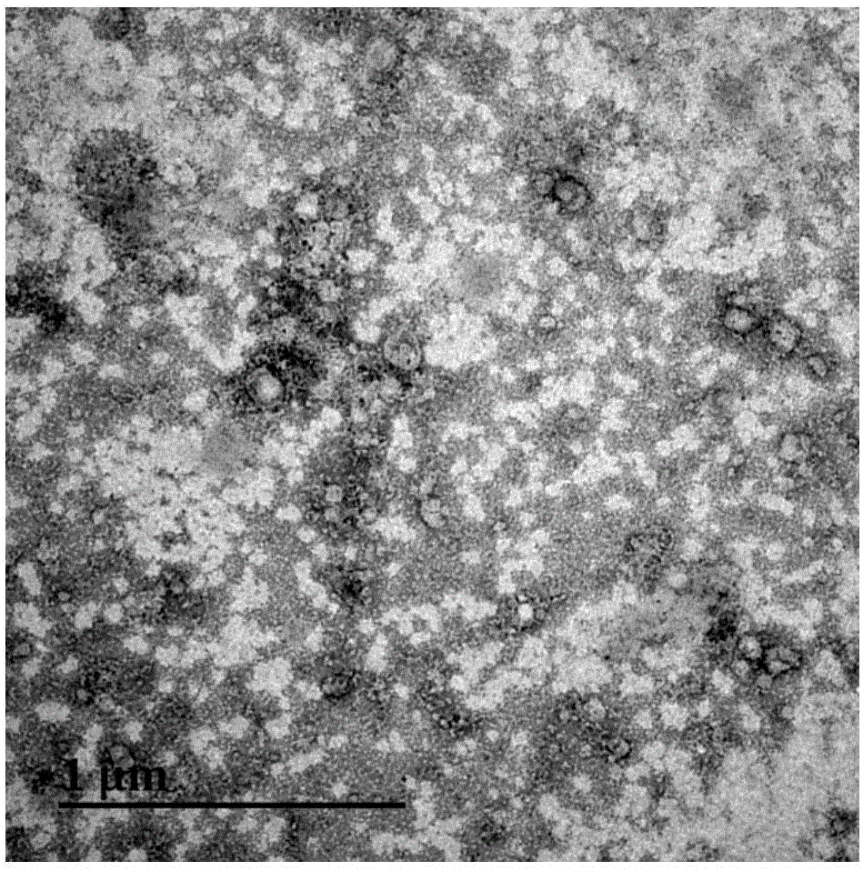
[0053] Example 3 Extraction of Exosomes (50-150nm) Subclasses in Microvesicles

[0054] (1) will 1*10 6 Individual gastric cancer cells (undifferentiated) HGC-27 were seeded at 75cm 2 Add RPMI-1640 culture medium containing 10% fetal bovine serum to the cell bottle, and place at 37°C, 5% CO 2 Cultivate in an incubator for 24 hours, collect 10ml of cell culture solution, add it to the first centrifuge tube, centrifuge at 200×g for 10 minutes, and separate to obtain 9.5ml of supernatant A;

[0055] (2) Add 9.5ml supernatant A to the second centrifuge tube, centrifuge at 2000×g for 40 minutes to obtain 9.0ml supernatant B;

[0056] (3) Add 9.0ml supernatant B to the third centrifuge tube, centrifuge at 10000×g for 40 minutes to obtain 8.5ml supernatant C;

[0057] (4) Add 8.5ml of supernatant C into a 100KDa ultrafiltration tube, and centrifuge at 5000rpm for 15 minutes. The filtrate was discarded, and the remaining liquid in the ultrafiltration tube was collected in a third ...


PUM


Abstract

The invention discloses a method for separating MVs (microvesicles) and MV exosomes from a tumor cell supernatant. The method comprises the following steps: after adherent culture of tumor cells, a culture solution is taken and subjected to centrifugation, the supernatant is mixed with a coupling compound, the mixture is incubated at 4 DEG C under the 100-300-rpm horizontal vibration condition for 3-16 h, a liquid is removed, an incubated coupling compound is obtained and cleaned with PBS (phosphate buffered saline), and coupling microspheres with MVs adsorbed are obtained. The total MVs extracted with the method retain the structures and components of all the MVs, and the yield is high. Secondary sorting can be performed, specific MVs can be analyzed, and the extraction efficiency and the research value are increased. The total extraction time requires only at least 4 hours to finish purification of the MVs and requires only at least 5 hours to finish purification of the exosomes. The extraction purity is higher, and experiments show that the extracted MVs account for 90% or higher of the total extracted proteins while organic precipitate accounts for lower than 20%.

Description

[0001] (1) Technical field [0002] The invention relates to the separation of microvesicles, in particular to a method for isolating tumor cell-derived microvesicles and exosomes from cell supernatants. [0003] (2) Background technology [0004] Microvesicles (MVs) are membranous structures shed or released from the cell membrane, and are membranous vesicles with a bilayer. MVs contain lipids, proteins, mRNA, microRNA (miRNA) and DNA fragments, etc., and these substances originate from the mother cell membrane or organelles. Not only in cells cultured in vitro but also in body fluids. Current studies have found that it plays a role in transmitting information between cells, tissues, and organs, and can regulate target cells, tissues, and organs through the biomolecules on its surface or contained in it. It not only plays an important role in the occurrence and development of various diseases, but also has a close relationship with tumor invasion, metastasis and colonization...

Claims


Application Information

Patent Timeline
IPC IPC(8): G01N1/34G01N1/30
CPCG01N1/30G01N1/34
Inventor 程向东吕航徐志远
Owner ZHEJIANG PROVINCIAL HOSPITAL OF TRADITIONAL CHINESE MEDICINE
Why Eureka
Try Eureka