Biological probe for detecting surface tension change of living cell membrane

A biological probe and surface tension technology, applied in the fields of cell biology and molecular biology, can solve the problems of high substrate material requirements and low spatial resolution, and achieve the effect of low cost and no side effects

Active Publication Date: 2017-02-15
DALIAN UNIV OF TECH
View PDF3 Cites 8 Cited by
  • Summary
  • Abstract
  • Description
  • Claims
  • Application Information

AI Technical Summary

Problems solved by technology

At present, the methods for detecting the surface tension of cell membranes include deformable polymer substrate method and microcolumn method, etc., but they have defects such as low spatial resolution and high requirements for substrate materials. Changes in tension are measured

Method used


Image

  • Biological probe for detecting surface tension change of living cell membrane
  • Biological probe for detecting surface tension change of living cell membrane
  • Biological probe for detecting surface tension change of living cell membrane

Examples


Embodiment Construction

[0023] The specific implementation manners of the present invention will be further described below in conjunction with the accompanying drawings and technical solutions.

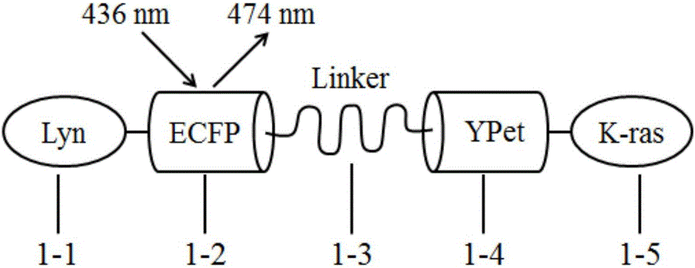
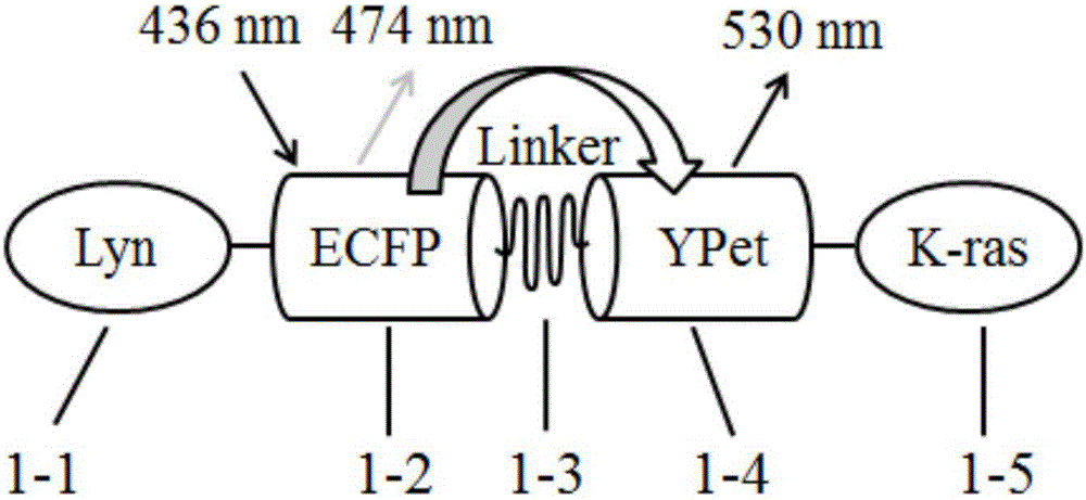
[0024] After the probe is transfected into living cells with a transfection reagent, it can express a fluorescent protein fusion probe structure by itself, and the Lyn sequences 1-1 and K-ras sequences 1-5 at both ends are respectively connected to the cell membrane. When the force changes the surface tension of the cell membrane, the elastic sequence Linker1-3 will be stretched or compressed, causing the FRET fluorescent protein to change the distance between ECFP 1-2 and YPet1-4, resulting in a change in the FRET signal. The transfected cells were excited with a wavelength of 436nm, and the fluorescence images at the wavelengths of 474nm and 530nm were collected simultaneously by FRET microscope, and then the efficiency of energy transfer was analyzed by the ratio of 474nm / 530nm fluorescence intensity, so ...


PUM


Abstract

The invention belongs to the technical fields of cell biology and molecular biology, and relates to an FRET technology-based biological probe for dynamically detecting the surface tension change of a living cell membrane, and concretely discloses a biological probe for detecting the surface tension change of a living cell membrane. The probe comprises FRET fluorescin pairs ECFP and YPet, an elastic sequence Linker, a Lyn sequence and a K-ras sequence. A fluorescin fusion probe can be spontaneously expressed in a cell after the biological probe and a pcDNA3.1(+) recombinant plasmid are transferred to the living cell, can qualitatively detect the tension change of the cell membrane, and has no influences on the original functions of the cell. The probe provided by the invention realizes dynamical detection of the surface tension change of the cell membrane in living cells, has the characteristics of low cost and no toxic or side effects on cells, and provides a visual tool for researches of the tension change of the cell membrane.

Description

technical field [0001] The invention belongs to the technical field of cell biology and molecular biology, and is a biological probe designed and prepared based on fluorescence resonance energy transfer (FRET) technology and subcloning technology. It can express itself in living cells and qualitatively reflect changes in cell membrane surface tension through changes in fluorescent signals. Background technique [0002] The shear stress acts on the upper surface of the cell membrane, which first causes a change in the surface tension of the cell membrane. This physical change may be the basis for the directional migration of tumor cells. At present, the methods for detecting the surface tension of cell membranes include deformable polymer substrate method and microcolumn method, etc., but they have defects such as low spatial resolution and high requirements for substrate materials. Changes in tension are measured. Therefore, the present invention proposes a biological prob...

Claims


Application Information

Patent Timeline
Patent Type & Authority Applications(China)
IPC IPC(8): C12N15/62C07K19/00C12Q1/02G01N21/64
CPCC07K14/43595C07K2319/00G01N21/6486G01N33/56966G01N33/68G01N2333/47
Inventor 刘波李旺于新磊张树利孙坚原
Owner DALIAN UNIV OF TECH
Why Eureka
Try Eureka