Method and device for acquiring cell agglutination graph

An acquisition method and cell technology, applied in the field of medical devices, can solve the problems such as the inability to record the non-linear characteristics of erythrocyte sedimentation velocity, the inability to meet the requirements of modern precision medicine, and the unintuitive monitoring results, achieving light weight, compact structure, and three-dimensional sense. strong effect

Pending Publication Date: 2017-03-22
山东朗伯光谱设备有限公司
View PDF6 Cites 3 Cited by
  • Summary
  • Abstract
  • Description
  • Claims
  • Application Information

AI Technical Summary

Problems solved by technology

[0008] 1. The monitoring results are not intuitive enough to meet the requirements of modern precision medicine;
[0009] 2. The transmission mechanism is not stable;
[0010] 3. It is impossible to remember the nonlinear characteristics of the red blood cell sedimen...

Method used


Image

  • Method and device for acquiring cell agglutination graph
  • Method and device for acquiring cell agglutination graph
  • Method and device for acquiring cell agglutination graph

Examples


Embodiment 1

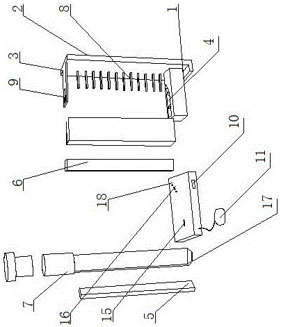
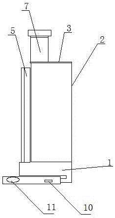
[0065] A cell agglutination map acquisition device, including a bracket with a mountain-shaped cross section, a CIS sensor and a base 18;

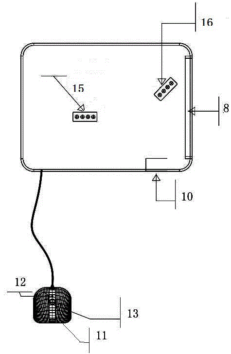
[0066] The bracket includes a positioning seat 1, a side wall 2 and a top wall 3; the CIS sensor includes a reflective light guide column 6, a transmissive light guide column 5, a reflective point light source 16, a transmissive point light source 15 and a CIS receiving module 8; a reflective light guide column 6, a transmissive light guide column Both 5 and the CIS receiving module 8 are arranged vertically to the positioning seat 1 .

[0067] The positioning seat 1 of the bracket is equipped with a test tube positioning socket 4, and the ESR tube 7 is inserted into the test tube positioning socket 4. The two sides of the ESR tube 7 are respectively equipped with a transmission light guide column 5 and a reflection light guide column 6, and the transmission light guide column 5 and the ESR tube 7 are respectively arranged. The front side ...

Embodiment 2

[0074] A cell agglutination map acquisition device, including a bracket, a CIS sensor and a base 18;

[0075] The bracket includes a positioning seat 1 and a side wall 2; the CIS sensor includes a transmission light guide column 5, a transmission point light source 15 and a CIS receiving module 8; the transmission light guide column 5 and the CIS receiving module 8 are arranged perpendicular to the positioning seat 1;

[0076] The positioning seat 1 of the bracket is equipped with a test tube positioning socket 4, the ESR tube 7 is inserted into the test tube positioning socket 4, and one side of the ESR tube 7 is equipped with a transmission light guide column 5, and the transmission light guide column 5 is closely attached to the side wall of the ESR tube 7, The ESR tube 7 is located between the transmission light guide column 5 and the CIS receiving module 8, the transmission light guide column 5 is facing the center of the ESR tube 7, and the CIS receiving module 8 is insta...

Embodiment 3

[0081] A cell agglutination map acquisition device, including a bracket with a mountain-shaped cross section, a CIS sensor and a base 18;

[0082] The bracket includes a positioning seat 1, a side wall 2 and a top wall 3; the CIS sensor includes a reflected line light source and a CIS receiving module 8;

[0083] The positioning seat 1 of the bracket is equipped with a test tube positioning socket 4, and the ESR tube 7 is vertically inserted into the test tube positioning socket 4, and one side of the ESR tube 7 is equipped with a reflected line light source, which is perpendicular to the positioning seat 1, and the reflected line light source and the ESR tube The side wall of 7 is arranged at 45 degrees, and the reflected line light source is located between the ESR tube 7 and the CIS receiving module 8, and the CIS receiving module 8 is installed on the side wall 2;

[0084] The reflected line light source runs through the positioning seat 1 and is connected to the base 18 ;...


PUM


Abstract

The invention discloses a method and device for acquiring a cell agglutination graph. The method specifically includes the steps of S1, a CIS sensor comprises a ray-transmitted light source and a CIS receiving module, and the ray-transmitted light source transmits a light signal; S2, the light signal vertically irradiates towards a blood sedimentation tube which is located between the ray-transmitted light source and the CIS receiving module; S3, the light signal is transmitted by the blood sedimentation tube and received by the CIS receiving module; S4, the CIS sensor works every other fixed seconds; S5, the light intensity of the transmitted light received by the CIS receiving module is different along with the blood sedimentation condition change in the blood sedimentation tube, the CIS receiving module converts the light signal into an electric signal, the electric signal is transmitted to a computer, and the computer generates the cell agglutination graph through calculation, parameter analysis and vivid simulation. The method has the advantages that the cell agglutination graph is generated through the simulation, blood sedimentation conditions can be displayed accurately, visually and dynamically, the sedimentation conditions of red blood cells are clear at a glance, doctors can performs real-time analysis conveniently, and the method is suitable for precision medicine; the method is applicable to the labs of various medical institutions and even applicable to families.

Description

technical field [0001] The invention relates to the technical field of medical devices, in particular to a method and device for obtaining a cell agglutination map. Background technique [0002] ESR is a common name for "erythrocyte sedimentation rate". Put the anticoagulated blood into the ESR tube and let it stand vertically, and the red blood cells will sink due to their high density. The sedimentation speed of red blood cells is usually expressed by the distance that red blood cells sink at the end of the first hour, which is called erythrocyte sedimentation rate, or erythrocyte sedimentation rate. [0003] The speed of erythrocyte sedimentation is related to plasma viscosity, especially the aggregation force between red blood cells. The higher the aggregation force between red blood cells, the faster the erythrocyte sedimentation rate, and vice versa. [0004] The determination of erythrocyte sedimentation rate is widely used clinically, such as acute glomerulonephri...

Claims


Application Information

Patent Timeline
IPC IPC(8): G01N15/05
CPCG01N15/05
Inventor 滕浩然肖昌林张攀
Owner 山东朗伯光谱设备有限公司
Why Eureka
Try Eureka