Micro-fluidic chip-based multiple-organ tumor targeting drug testing platform and application thereof
A technology of microfluidic chip and testing platform, which is applied to the measurement/inspection of microorganisms, the method of supporting/immobilizing microorganisms, biochemical instruments, etc., and can solve problems such as being in the blank stage
- Summary
- Abstract
- Description
- Claims
- Application Information
AI Technical Summary
Problems solved by technology
Method used
Image
Examples
Embodiment 1
[0026] Establishment of multi-organ tumor targeting drug testing platform chip
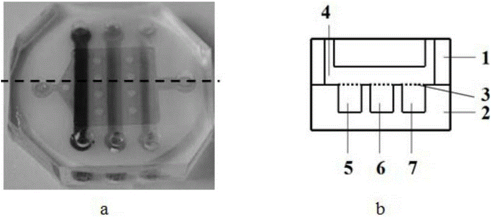
[0027] Design and manufacture chips, the upper chip 1 is a cell culture chamber 4, the lower chip 2 is three parallel independent channels, the structure is as follows figure 1 as shown in b. The porous filter membrane 3 is placed on a glass slide for UV activation for 1 hour, silanized for 30 minutes, sealed with the upper chip 1 by oxygen plasma, and placed in an oven at 80 degrees for 30 minutes. Use a PDMS polymer with a monomer-to-initiator ratio of 20:1, and throw it on a glass slide to a thickness of 10um-50um. After dipping the upper surface of the lower chip 2 into thin PDMS, it is aligned and bonded to the porous filter membrane 3 sealed with the upper chip. , 80 degrees, 30 minutes to cure completely. The result is as figure 1 as shown in a.
Embodiment 2
[0029] Characterization of a multi-organ tumor-targeted drug testing platform
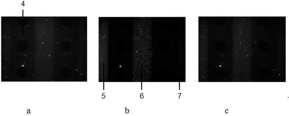
[0030] The microfluidic chip designed and manufactured by the laboratory itself has a structure such as figure 1 as shown in b. After Hepg2 cells were treated with green cell membrane dye for 30 min, they were inserted into cell culture chamber 4 on the upper chip. After MDA-MB-231 cells, A549 cells, and GES-1 cells were treated with red cell membrane dye for 30 minutes, they were respectively inserted into the three channels of the lower chip, 1# channel 5, 2# channel 6, and 3# channel 7. Use a fluorescence microscope to photograph the cells in the cell culture chamber 4 on the upper layer of the chip, and the results are as follows figure 2 As shown in a, the cells of channel 1# channel 5, channel 2# channel 6, and channel 3# 7 of the lower chip channel were photographed with a fluorescent microscope, and the results are as follows figure 2 as shown in b. Merge the fluorescence image of the...
Embodiment 3
[0032] Testing the effect of capecitabine on different cells using a multi-organ tumor-targeted drug testing platform
[0033] Using the microfluidic chip designed and manufactured by the laboratory, the structure is as follows: figure 1 as shown in b. Prepare a type I mouse tail collagen working solution with a concentration of 100 μg / mL, inject it into the chip through the cell inlet pool, let it stand at room temperature overnight, remove the collagen working solution after 24 hours, and wash the channel 3 times with the cell culture solution. When the upper chip was inoculated / not inoculated with Hepg2 cells, the three units of the lower chip were inoculated with MDA-MB-231, U87, and GES-1 cells, and H-DMEM medium containing 80 μM capecitabine was added to the upper culture chamber. After 48 hours, the toxic effect of the drug on different cells was detected by a dead / alive staining kit and a CCK8 kit. The result is as image 3 a and image 3 As shown in b, when capeci...
PUM
 Abstract
Description
Claims
Application Information
 - R&D
- Intellectual Property
- Life Sciences
- Materials
- Tech Scout
- Unparalleled Data Quality
- Higher Quality Content
- 60% Fewer Hallucinations
Browse by: Latest US Patents, China's latest patents, Technical Efficacy Thesaurus, Application Domain, Technology Topic, Popular Technical Reports.
© 2025 PatSnap. All rights reserved.Legal|Privacy policy|Modern Slavery Act Transparency Statement|Sitemap|About US| Contact US: help@patsnap.com


