Method for segmenting endocardium and epicardium in heart cardiac function magnetic resonance image

A technology for magnetic resonance images and cardiac function, applied in the field of medical images, can solve the problems of complicated, insufficiently accurate extraction of the left ventricular epicardium, and complicated semi-automatic detection methods, so as to achieve the effect of improving efficiency and accuracy

Active Publication Date: 2017-06-30
SHANGHAI UNITED IMAGING HEALTHCARE
View PDF7 Cites 8 Cited by
  • Summary
  • Abstract
  • Description
  • Claims
  • Application Information

AI Technical Summary

Problems solved by technology

Although many research methods and results have been reported in the past 20 years, the accurate detection of left ventricular endocardium in four-dimensional functional magnetic resonance imaging is still unresolved.
For the detection of left ventricular endocardium in four-dimensional cardiac functional magnetic resonance images, manual and user-assisted semi-automatic detection methods are complicated, inefficient, and there are large differences between observers and among observers
In terms of fully automatic detection, due to the huge differences in grayscale and gradient distribution of four-dimensional functional magnetic resonance images in different patients, different scanning tomograms of the same patient, and the same scanning tomographic images, in addition, left ventricular epicardium The diversity of surrounding adjacent tissues makes simple threshold-based detection methods or gradient-based detection methods insufficient to accurately extract the endocardium of the left ventricle
Model-based detection methods require a large number of training samples to train the model to obtain more accurate detection results, and the acquisition of these accurate training samples is a more difficult and complicated task

Method used


Image

  • Method for segmenting endocardium and epicardium in heart cardiac function magnetic resonance image
  • Method for segmenting endocardium and epicardium in heart cardiac function magnetic resonance image
  • Method for segmenting endocardium and epicardium in heart cardiac function magnetic resonance image

Examples


Embodiment Construction

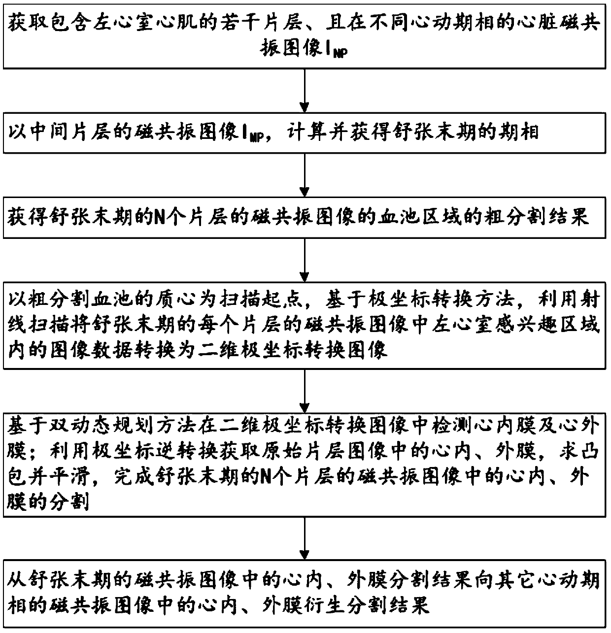
[0044] see Figure 1-5 , a method for segmenting endocardium and endocardium in a functional magnetic resonance image of the heart in an embodiment of the present invention, is characterized in that comprising the following steps:

[0045] S1. Acquire cardiac magnetic resonance images including several slices of left ventricular myocardium and in different cardiac phases I NP , where N represents the sequence number of the slice, P represents the sequence number of the cardiac phase, and both N and P are integers greater than or equal to 1;

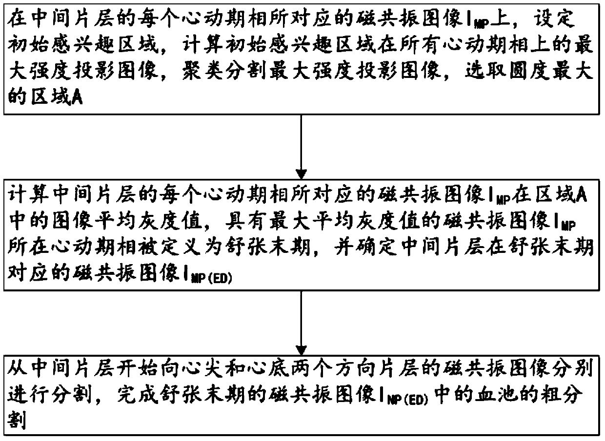
[0046] S2. Determining the phase of end diastole;

[0047] S3. Obtain a rough segmentation result of the blood pool area of ​​the magnetic resonance image of N slices at the end of diastole;

[0048] S4. Taking the centroid of the roughly segmented blood pool as the scanning starting point, based on the polar coordinate conversion method, the image data in the left ventricle region of interest in the magnetic resonance image of each sli...


PUM


Abstract

The invention discloses a method for segmenting the endocardium and the epicardium in a heart cardiac function magnetic resonance image. The method comprises positioning the diastasis in heart magnetic resonance images I<NP> including several lamellas of the left ventricular myocardium and in different cardiac cycle phases, and obtaining a coarse segmentation result of blood pools of magnetic resonance images of N lamellas in the diastasis; converting image data in the left ventricle region-of-interest in the magnetic resonance image of each lamella in the diastasis into a two-dimension polar coordinate conversion image by means of ray scanning based on a polar coordinate conversion method; detecting the endocardium and the epicardium in the two-dimension polar coordinate conversion image based on a bidynamic programming method; obtaining the endocardium and the epicardium in an original lamella image by means of polar coordinate inverse conversion, calculating the convex hull and smoothing the convex hull, and completing segmentation of the endocardium and the epicardium in the magnetic resonance images of the N lamellas in the diastasis; and deriving the segmentation result of the in the endocardium and the epicardium in the magnetic resonance images in the diastasis to the endocardium and the epicardium in the magnetic resonance images in other cardiac cycle phases.

Description

【Technical field】 [0001] The invention relates to the field of medical images, in particular to the segmentation of cardiac functional magnetic resonance images. 【Background technique】 [0002] In recent years, the morbidity and mortality of cardiovascular diseases are increasing year by year. Nearly 6 million people in the United States are diagnosed with cardiovascular disease every year, while nearly 3 million people die of cardiovascular disease in my country. Cardiac function is an important indicator for the diagnosis of cardiovascular diseases, which can be achieved by analyzing the global and local characteristic parameters related to the left ventricle in the four-dimensional functional magnetic resonance image, such as: ventricular volume, ejection fraction, and myocardial wall thickness. The accuracy of the extraction of cardiac function feature parameters depends on the accurate localization of the left ventricular endocardium, and the computer-aided diagnosis s...

Claims


Application Information

Patent Timeline
IPC IPC(8): G06T7/11G06F19/00
CPCG06T2207/10088G06T2207/30048
Inventor 姜娈凌姗李强
Owner SHANGHAI UNITED IMAGING HEALTHCARE
Why Eureka
Try Eureka