Segmentation method of epicardium and epicardium in cardiac functional magnetic resonance images
A magnetic resonance image and heart function technology, applied in the field of medical images, can solve problems such as complexity, low efficiency, and unsolved detection, and achieve the effect of improving efficiency and accuracy
- Summary
- Abstract
- Description
- Claims
- Application Information
AI Technical Summary
Problems solved by technology
Method used
Image
Examples
Embodiment Construction
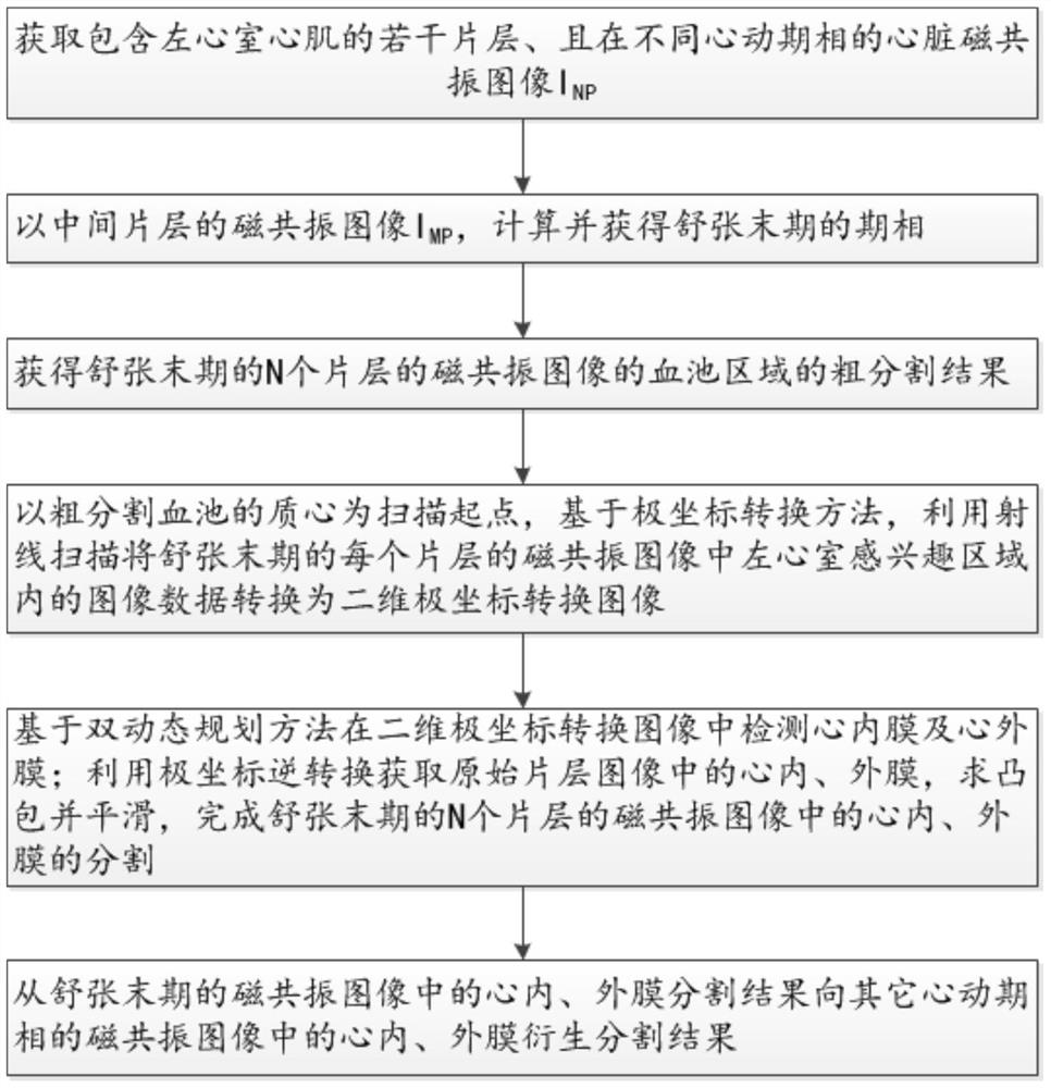
[0044] see Figure 1-5 , a method for segmenting endocardium and endocardium in a functional magnetic resonance image of the heart in an embodiment of the present invention, is characterized in that comprising the following steps:
[0045] S1. Acquire cardiac magnetic resonance images including several slices of left ventricular myocardium and in different cardiac phases I NP , where N represents the sequence number of the slice, P represents the sequence number of the cardiac phase, and both N and P are integers greater than or equal to 1;
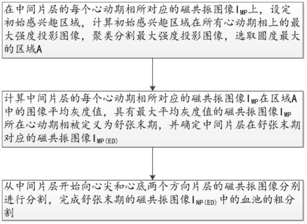
[0046] S2. Determining the phase of end diastole;
[0047] S3. Obtain a rough segmentation result of the blood pool area of the magnetic resonance image of N slices at the end of diastole;
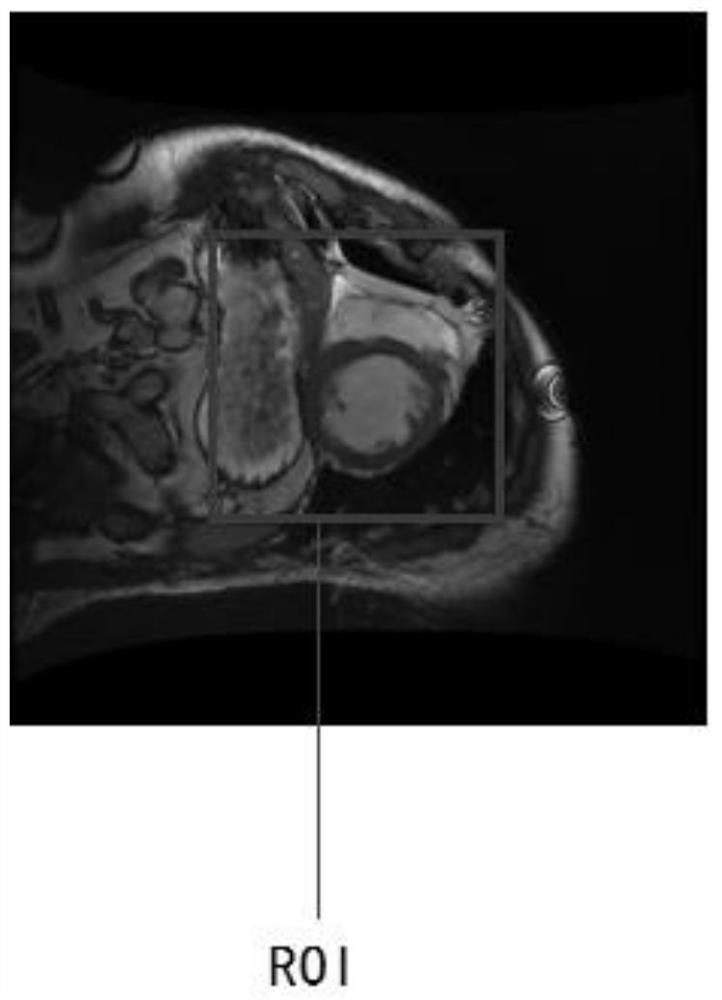
[0048] S4. Taking the centroid of the roughly segmented blood pool as the scanning starting point, based on the polar coordinate conversion method, the image data in the left ventricle region of interest in the magnetic resonance image of each sli...
PUM
 Abstract
Description
Claims
Application Information
 - R&D
- Intellectual Property
- Life Sciences
- Materials
- Tech Scout
- Unparalleled Data Quality
- Higher Quality Content
- 60% Fewer Hallucinations
Browse by: Latest US Patents, China's latest patents, Technical Efficacy Thesaurus, Application Domain, Technology Topic, Popular Technical Reports.
© 2025 PatSnap. All rights reserved.Legal|Privacy policy|Modern Slavery Act Transparency Statement|Sitemap|About US| Contact US: help@patsnap.com


