Method for constructing three-dimensional model based on tissue slices

A technology of tissue slices and three-dimensional models, applied in the field of biochemistry, can solve the problems of easy deformation of tissues, inability to display the three-dimensional shape and distribution of antigens, expensive experimental equipment, etc., and achieve the effect of low application threshold

Inactive Publication Date: 2017-07-04
WUHAN RUIFUNING TECH
View PDF6 Cites 4 Cited by
  • Summary
  • Abstract
  • Description
  • Claims
  • Application Information

AI Technical Summary

Problems solved by technology

Although conventional IHC techniques can present clear two-dimensional morphology and distribution of tissue antigens, they cannot reveal the three-dimensional morphology and distribution of antigens in tissues
The existing tissue 3D technology also has the following disadvantages: 1. The first tissue block transparent scanning technology has the following defects: it takes a long time (about 1-2 months), the tissue is easily deformed, and the transparent effect of large tissue blocks is poor; 2. Two-photon tomography Scanning technology and fluorescent micro-optical section tomography technology can only target tissues with autofluorescent labels, and cannot label tissue antigens through antibodies; 3. The realization of the above 3D technologies requires expensive experimental equipment

Method used


Image

  • Method for constructing three-dimensional model based on tissue slices
  • Method for constructing three-dimensional model based on tissue slices
  • Method for constructing three-dimensional model based on tissue slices

Examples


Embodiment 1

[0031] In this embodiment, the bright-field 3D reconstruction of cholinergic neurons in mouse brain tissue is taken as an example for illustration.

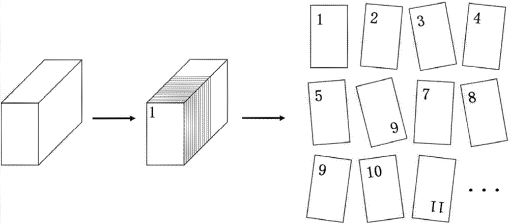
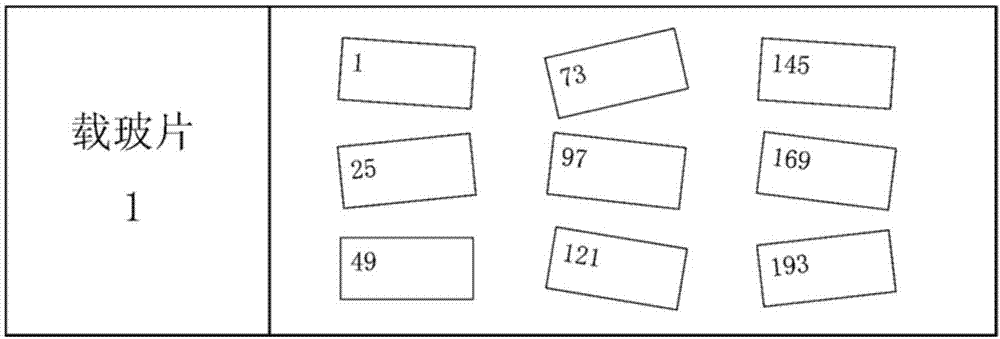
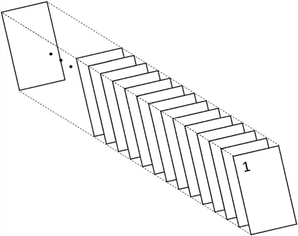
[0032] In the first step, the mouse brain tissue was first sliced ​​serially with a frozen microtome, and the slice thickness was 16 microns, and divided into 24 equal parts. The specific steps were as follows (such as figure 1 as shown, figure 1 It is a structural schematic diagram of continuous sectioning of animal tissue blocks in an embodiment of the present invention. ):

[0033] 1. Set the parameters of the cryostat (Leica, Germany) (the temperature of the fixed base is 16°C, the temperature of the chassis is 20°C, and the slice thickness is 16 microns), and install a brand-new blade (Leica, Germany), and then wait for 1 hour ;

[0034] 2. Coat the surface of the microtome base with frozen section embedding agent (Sakura, Japan), about 1 mm thick, and use a microtome to flatten the surface after it solidifies;

[0035] ...

Embodiment 2

[0071] In this example, the fluorescent 3D reconstruction of somatostatin-positive neurons in mouse brain tissue is taken as an example for illustration.

[0072] In the first step, the mouse brain tissue was serially sliced ​​with a frozen microtome with a thickness of 16 microns and divided into 24 equal parts. The specific steps are as follows:

[0073] 1. Set the parameters of the cryostat (Leica, Germany) (the temperature of the fixed base is 16°C, the temperature of the chassis is 20°C, and the slice thickness is 16 microns), and install a brand-new blade (Leica, Germany), and then wait for 1 hour ;

[0074] 2. Coat the surface of the microtome seat with tissue embedding agent (Sakura, Japan), about 1 mm thick, and use a microtome to flatten the surface after it solidifies;

[0075] 3. Cut the mouse brain tissue into two equal parts along the sagittal midline, and then poke two small holes in the brain tissue perpendicular to the right brain (or left brain) section (for...

Embodiment 3

[0099] In this example, the fluorescent 3D reconstruction of excitatory neurons in the brain tissue of transgenic mice is taken as an example for illustration.

[0100] In the first step, the mouse brain tissue was serially sliced ​​with a frozen microtome with a thickness of 16 microns and divided into 24 equal parts. The specific steps are as follows:

[0101] 1. Set the parameters of the cryostat (Leica, Germany) (the temperature of the fixed base is 16°C, the temperature of the chassis is 20°C, and the slice thickness is 16 microns), and install a brand-new blade (Leica, Germany), and then wait for 1 hour ;

[0102] 2. Coat the surface of the microtome seat with tissue embedding agent (Sakura, Japan), about 1 mm thick, and use a microtome to flatten the surface after it solidifies;

[0103] 3. Cut the mouse brain tissue into two equal parts along the sagittal midline, and then poke two small holes in the brain tissue perpendicular to the right brain (or left brain) sectio...


PUM

PropertyMeasurementUnit
diameteraaaaaaaaaa
thicknessaaaaaaaaaa
thicknessaaaaaaaaaa

Abstract

The invention discloses a method for constructing a three-dimensional model based on tissue slices. The method comprises 1, continuously slicing tissue to obtain several parts, carrying out dyeing labeling on all the tissue slices and completely pasting the tissue slices to a corresponding glass slide, or 2, slicing tissue to obtain tissue slices with fluorescence signals and completely pasting the tissue slices to a corresponding glass slide without labeling, and 3, orderly naming the tissue slices according to glass slide numbers through the scanned tissue slice images so that all the images can be arranged in a slice original order, carrying out calibration, and carrying out 3D synthesis on the calibrated images to obtain the three-dimensional model based on tissue slices. The method can realize flexible labeling of the conventional IHC technology and can be used as a conventional technology. The 3D-IHC can realizes antigen labeling of the conventional IHC technology.

Description

technical field [0001] The invention belongs to the technical field of biochemistry, and in particular relates to a method for constructing a three-dimensional model based on tissue slices. Background technique [0002] Immunohistochemistry (IHC, hereinafter referred to as IHC), also known as immunocytochemistry, refers to the reaction of specific antibodies labeled with chromogenic agents (fluorescein, enzymes, metal ions, isotopes) in situ on tissue cells through antigen-antibody reactions and tissue Chemical color reaction, a new technology for qualitative, localization and quantitative determination of corresponding antigens. It skillfully combines the specificity of the immune response and the visibility of histochemistry, and detects various antigenic substances (such as proteins, peptides, enzymes, hormones, pathogens and receptors, etc.). [0003] With the advancement of microscopic imaging technology, the following three three-dimensional (3D) histomorphological t...

Claims


Application Information

Patent Timeline
Patent Type & Authority Applications(China)
IPC IPC(8): G01N1/28
CPCG01N1/2813G01N2001/282
Inventor 颜欢欢庞培李浩吴卓泽鲁友明
Owner WUHAN RUIFUNING TECH
Why Eureka
Try Eureka