Method and probe for detecting miRNA (Ribonucleic Acid) and/or target molecule with aptamer

A nucleic acid aptamer and probe technology, applied in biochemical equipment and methods, DNA/RNA fragments, DNA preparation, etc., can solve problems such as high cost and complicated operation

Active Publication Date: 2017-07-18
LINYI UNIVERSITY
View PDF2 Cites 10 Cited by
  • Summary
  • Abstract
  • Description
  • Claims
  • Application Information

AI Technical Summary

Problems solved by technology

[0005] At present, there have been reports on the detection of ATP using biosensors based on ATP nucleic acid aptamers. Changes are detected, so the operation is complicated and the

Method used


Image

  • Method and probe for detecting miRNA (Ribonucleic Acid) and/or target molecule with aptamer
  • Method and probe for detecting miRNA (Ribonucleic Acid) and/or target molecule with aptamer
  • Method and probe for detecting miRNA (Ribonucleic Acid) and/or target molecule with aptamer

Examples


Embodiment 1

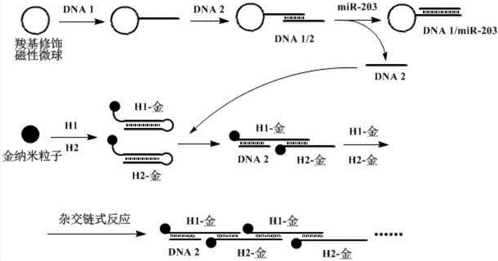
[0064] Embodiment 1: Detect the preparation of miRNA probe:

[0065] The probe includes: gold nanoparticle-H1 complex, gold nanoparticle-H2 complex and carboxyl magnetic microsphere-DNA1 / 2 complex; wherein: the sequences of H1 and H2 are as follows:

[0066] H1: 5'-SH-GCG ATT CCT AGG TTG AGC CCA GGG TTT TTT CACAGT CCC TGG GCTCAA CCT AGG-3'; (SEQ ID NO.1)

[0067] H2: 5'-CCC TGG GCT CAA CCT AGG AAT CGC TTT TTT CCT AGG TTGAGC CCA GGGACT GTG-SH-3'; (SEQ ID NO. 2).

[0068] DNA1 contains a sequence complementary and / or partially complementary to the miRNA to be detected; DNA1 is partially complementary to DNA2.

[0069] Wherein, the preparation method of gold nanoparticle-H1 composite, gold nanoparticle-H2 composite is:

[0070] (1) Preparation of gold nanoparticles with a diameter of 5nm: Add 0.6mL of 0.1M sodium borohydride on ice to 20mL of chloroauric acid (0.25mM) and trisodium citrate (0.25mM) mixture, and stir until the solution becomes Pink, reaction 2-5h. The gold nan...

Embodiment 2

[0074] Embodiment 2: the application of the probe of the present invention in detecting miR-203

[0075] 1. Test material:

[0076] The nucleotide sequence of miR-203 is as follows:

[0077] 5'-GUG AAA UGU UUA GGA CCA CUA G-3'.

[0078] The composition and preparation method of the probe are the same as in Example 1, in the probe:

[0079] The sequences of H1 and H2 are as follows:

[0080] H1: 5'-SH-GCG ATT CCT AGG TTG AGC CCA GGG TTT TTT CACAGT CCC TGG GCTCAA CCT AGG-3'; (SEQ ID NO.1)

[0081] H2: 5'-CCC TGG GCT CAA CCT AGG AAT CGC TTT TTT CCT AGG TTGAGC CCA GGGACT GTG-SH-3'; (SEQ ID NO.2);

[0082] The sequences of DNA1 and DNA2 are as follows:

[0083] DNA1: 5'-GGG C TAG TGG TCC TAA ACA TTT CAC-NH 2 -3'; (SEQ ID NO.3)

[0084] DNA2: 5'-GGA CCA CTA G CCC TGG GCT CAA CCT AGG AAT CGC-3'; (SEQ ID NO. 4).

[0085] 2. Detection method:

[0086] The specific method is as follows:

[0087] (1) Preparation of miR-203 working solution: Preparation of miR-203 working solution...

Embodiment 3

[0091] Example 3: Selectivity investigation of miR-203 detection method

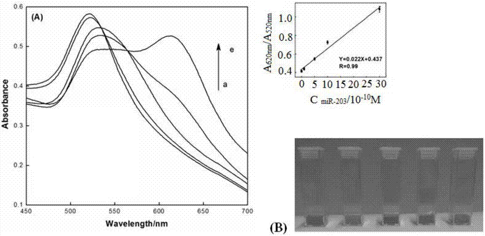
[0092] miR-203, miR-21 (nucleotide sequence: 5'-UAG CUU AUC AGA CUG AUG UUG A-3') and miR-16 (nucleotide sequence: 5'-UAG CAG CAC GUA AAU AUU GGC G-3') solution Preparation: Sterilize the PBS buffer solution (pH 7.4) containing 1‰ (volume ratio) DEPC water, and use it to prepare 1.0×10 -10 M miR-203, 2.0×10 -9 MmiR-21 and 2.0×10 -9 M miR-16 solution.

[0093] Mix 100 μL of carboxylated magnetic microsphere-DNA1 / 2 complex with 100 μL of miR-203 solution (1.0 × 10 -10 M) or miR-21 solution (1.0×10 -9 M) or miR-16 solution (1.0×10 -9 M), mix evenly, perform magnetic separation after reacting at 25°C for 2 hours, and absorb the supernatant, then add 100 μL gold nanoparticle-H1 complex and 100 μL gold nanoparticle-H2 complex to the supernatant, and incubate for 4 hours Finally, record the UV absorption peak ratio at 620nm and 520nm. Such as Figure 5 As shown, only the miR-203 solution caused signific...


PUM


Abstract

The invention discloses a method and probe for detecting miRNA (Ribonucleic Acid) and/or a target molecule with an aptamer. The probe comprises a gold nanoparticle-H1 compound, a gold nanoparticle-H2 compound and a target recognition compound, wherein the gold nanoparticle-H1 compound and the gold nanoparticle-H2 compound are respectively formed by bonding neck ring DNA (Deoxyribonucleic Acid) H1 and H2 to gold nanoparticles; and the target recognition compound comprises at least one of a carboxyl magnetic microsphere-DNA/2 compound for recognizing miRNA, and DNA3 for recognizing target molecules. The method disclosed by the invention does not need chemical markers, shows visible color change with naked eyes, is simple in operation and wide in application range and can be applied to detection on target molecules with the aptamer.

Description

technical field [0001] The invention relates to the technical field of nucleic acid hybridization detection, in particular to a detection method and a detection probe for miRNA and / or target molecules with nucleic acid aptamers. Background technique [0002] miRNA is a class of endogenous non-coding small molecules (18-22nt), which play an important regulatory role in gene expression or reverse transcription. It is associated with a wide range of biological processes such as cell proliferation, apoptosis and death. Aberrant expression of miRNAs has been implicated in various diseases, especially human cancers, neurological diseases, viral infections and diabetes. The obvious regulatory role of miRNA in physiological and pathological processes makes miRNA be used in the diagnosis of clinical diseases, gene therapy and the discovery of new anticancer drugs. For example, miRNA-203 is overexpressed in various malignancies, including breast cancer, cervical cancer, leukemia and...

Claims


Application Information

Patent Timeline
IPC IPC(8): C12Q1/68C12N15/11C12N15/10G01N33/573G01N33/68
CPCC12Q1/6816G01N33/573G01N33/5735G01N33/68C12Q2563/137C12Q2563/155C12Q2525/301C12Q2525/207C12Q2563/143C12Q2563/149
Inventor 郭英姝李双尚鑫鑫王玉洁张书圣
Owner LINYI UNIVERSITY
Why Eureka
Try Eureka