Method for quantitatively detecting glial fibrillary acidic protein (GFAP) in blood serum

A kind of neuroglial, quantitative detection technology, applied in the fields of nanomaterials, fluorescence immunology technology and biological analysis detection, to achieve the effect of reducing interference and good water solubility

Inactive Publication Date: 2017-08-29
NANJING MEDICAL UNIV
View PDF6 Cites 3 Cited by
  • Summary
  • Abstract
  • Description
  • Claims
  • Application Information

AI Technical Summary

Problems solved by technology

At present, the research mainly focuses on the combination of carbon dots and aptamers for the detection system, and the work of combining with antibodies for detection is still in its infancy, and there is no report on the application of CDs combined with antibodies to detect GFAP.

Method used


Image

  • Method for quantitatively detecting glial fibrillary acidic protein (GFAP) in blood serum
  • Method for quantitatively detecting glial fibrillary acidic protein (GFAP) in blood serum
  • Method for quantitatively detecting glial fibrillary acidic protein (GFAP) in blood serum

Examples


Embodiment 1

[0035] Example 1 Preparation of protein A / G agarose purified resin-linked antibody (PA / G-Ab1), the steps are as follows:

[0036] Add 10 μL of protein A / G agarose purification resin to a centrifuge tube, add 0.5 mL of binding buffer to resuspend the resin, centrifuge at 3000 rpm for 3 minutes, discard the supernatant, and repeat twice; add mouse anti-human GFAP to the balanced PA / G Monoclonal IgG 45μL (20μg / mL), 100rpm, shake and mix at 25°C for 1h; after the reaction, add 1% BSA and react for 1h to block the non-specific sites on the antibody; the reaction product PA / G-Ab1 was mixed with binding buffer Wash with liquid, centrifuge at 3000rpm for 3min, discard the supernatant, repeat twice to remove unreacted Ab1 and impurities, and store the product at 4°C until use.

Embodiment 2

[0037] The preparation of embodiment 2. carbon dots (CDs), the steps are as follows:

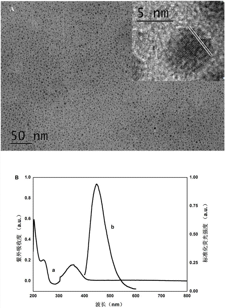
[0038] Take 1.2g of citric acid monohydrate, 1.2mL of diethylenetriamine and 20mL of ultrapure water in a 30mL autoclave, fully ultrasonicate for 15min to dissolve, and react at 200°C for 6h; take out the autoclave and place it at room temperature to obtain a brown solution. That is the CDs solution; in order to purify CDs, add phosphoric acid to adjust the pH to about 7, dialyze overnight with a 1000Da dialysis bag to remove unreacted impurities; collect the dialyzed CDs solution, suspend under reduced pressure, concentrate and dry at low temperature; the obtained CDs are pale yellow powders. Its transmission electron microscope image ( figure 1 A) UV absorption and fluorescence emission diagrams ( figure 1 B).

Embodiment 3

[0039] The preparation of embodiment 3 fluorescently labeled detection antibodies (CDs-Ab2), the steps are as follows:

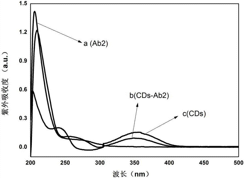
[0040] Dissolve CDs in 10mM PBS to make a 200μg / mL solution; add 30μL each of EDC and NHS (200mg / mL) to 150μL CDs, add NaOH to adjust the pH of the solution to about 7.5, and ultrasonically react for 30min; add rabbit anti-human GFAP Clone IgG, 200rpm, shaking reaction at 25°C for 2.5h; the reaction system was blocked with 1% BSA to block the non-specific site of the antibody, and reacted at 25°C for 1h; Fluorescence; the resulting product CDs-Ab2 (b) and Ab2 (a), CDs (c) were measured ultraviolet, the results are as follows figure 2 shown.


PUM

PropertyMeasurementUnit
particle diameteraaaaaaaaaa

Abstract

The invention discloses a method for quantitatively detecting glial fibrillary acidic protein (GFAP) in a blood serum. The method comprises the steps of firstly, purifying a resin connecting antibody Ab1 through protein A/G agarose, capturing GFAP (Glial Fibrillary Acidic Protein) in the blood serum to form PA/G-Ab1-GFAP, and centrifugally cleaning to achieve the aims of separation and enrichment; secondly, adopting citric acid as a carbon source and diethylenetriamine as a passivator for synthesizing carbon dots (CDs), connecting with an antibody Ab2 through an amido bond, and forming a fluorescence labelled probe CDs-Ab2; then combining the PA/G-Ab1-GFAP and the CDs-Ab2 to form a sandwich structure PA/G-Ab1-GFAP-Ab2-CDs, detecting fluorescence intensity, and obtaining the content of the GFAP in the blood serum according to a standard curve. The method provided by the invention has the advantages of high sensitivity and simplicity and convenience in detection, and the characteristics of high selectivity and high affinity of immunoreaction, and can be directly applied in determining the GFAP in the blood serum.

Description

technical field [0001] The invention belongs to the fields of nanomaterials, fluorescent immunology technology and biological analysis and detection, and in particular relates to a method for quantitatively measuring glial fibrillary acidic protein (GFAP) in serum by double-antibody sandwich based on carbon dots as fluorescent markers. Background technique [0002] Traumatic brain injury (TBI) is the leading cause of death in adolescents and children. Traumatic brain injury includes initial injury of different grades and the process of cell death centered on the point of initial injury and transmission of injury to surrounding tissues over time. Therefore, there is a need to establish a suitable method for detecting traumatic brain injury. Currently, the main methods for diagnosing traumatic brain injury are as follows: magnetic resonance imaging (MRT), computed tomography (CT), diffusion tensor imaging (DTI) and hospital observation. However, these existing diagnostic met...

Claims


Application Information

Patent Timeline
Patent Type & Authority Applications(China)
IPC IPC(8): G01N33/68G01N33/533
CPCG01N33/6893G01N33/533
Inventor 胡琴马云苏许贯虹魏芳弟岑瑶
Owner NANJING MEDICAL UNIV
Why Eureka
Try Eureka