

Cell culture fluid inducing umbilical cord mesenchymal stem cells into neural stem cells, and using method thereof
A cell culture, stem cell technology, applied in the direction of cell culture active agent, animal cells, culture process, etc., to achieve the effect of high proportion, shortened time and obvious characteristics
- Summary
- Abstract
- Description
- Claims
- Application Information
AI Technical Summary
Problems solved by technology
Method used
Image

Examples
Embodiment 1
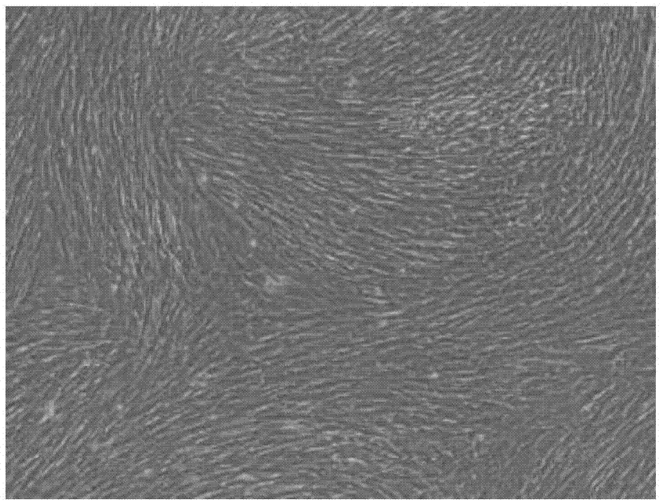
[0030] Take fresh healthy umbilical cord and umbilical cord blood, collect the umbilical cord blood and centrifuge, take the upper serum and inactivate it for later use. After rinsing with PBS, remove the umbilical vessels with scissors and tweezers, peel off the Wahrenheit jelly tissue inside, and cut the resulting tissue to a size of 1mm3, add α-MEM culture medium and place at 37°C, 5% CO 2 Cultivate in an incubator, and the culture medium contains 10% inactivated umbilical cord serum, 100U / ml streptomycin, and 100U / ml penicillin. After 5-8 days of umbilical cord tissue culture, it can be seen that some cells crawled out from around the tissue block, showing a small spindle shape. After one week, the cells began to proliferate rapidly and formed cell colonies of various sizes. After the cells were full, use 0.25 % Trypsinization for passage. Such as figure 1 As shown, the human umbilical cord mesenchymal stem cells grow in the form of fibroblast-like adherence, and the cel...
Embodiment 2
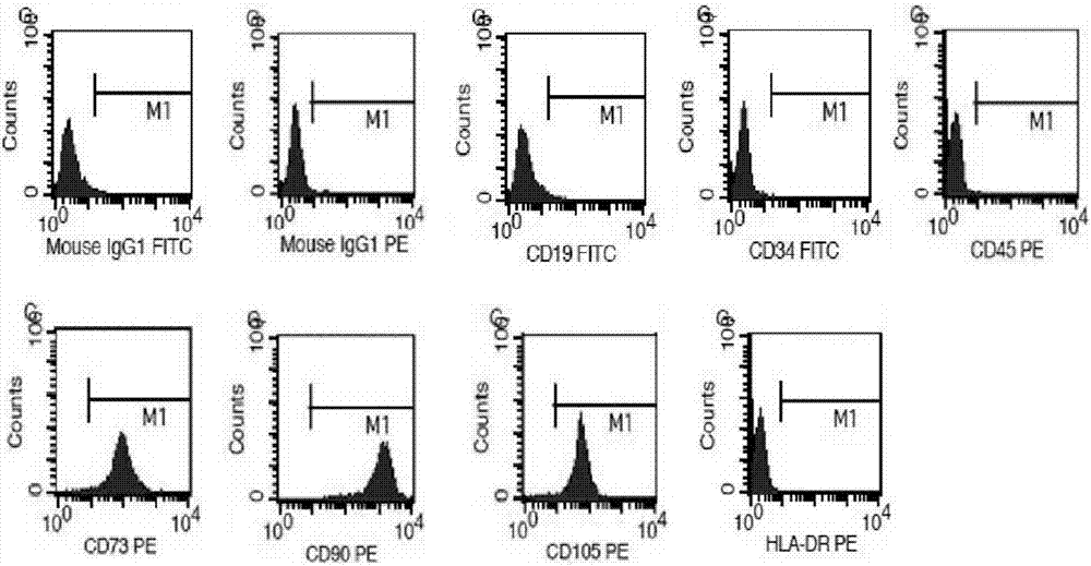
[0032] Take P3 UC-MSCs in the logarithmic growth phase, digest with trypsin, and adjust the cell density to 1x10 6 cells / ml, transfer the cells to 9 flow tubes, 1ml in each tube, centrifuge at 600g for 3min, discard the supernatant, add 1ml PBS to wash 2 times. After resuspending the cells in 200 μl PBS, add 5 μL each of CD19-FITC, CD34-FITC, CD45-PE, CD73-PE, CD90-PE, CD105-PE and HLA-DR-PE mouse anti-human monoclonal antibodies, and respectively Add 5 μL of anti-mouse IgG1-FITC and anti-mouse IgG1-PE to the other 2 tubes as isotype controls. After incubating at 4°C in the dark for 30 min, wash with PBS three times, discard the supernatant, resuspend the cells in PBS, and detect the cell phenotype by flow cytometry.
[0033] The result is as figure 2 As shown, the UC-MSCs obtained by isolation and culture fully meet the identification criteria, that is, high expression of CD73, CD90, and CD105, low expression or no expression of CD19, CD34, CD45, and HLA-DR.
Embodiment 3
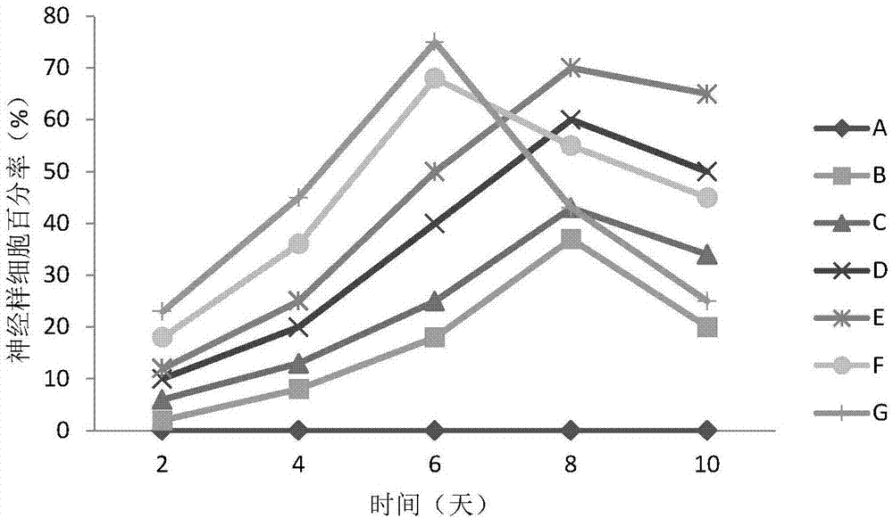
[0035] Take P3 UC-MSCs in the logarithmic growth phase, digest with trypsin, and adjust the cell density to 1x10 5 cells / ml, inoculated in 6-well plates, 2ml per well, replaced with fresh culture medium after 24 hours, and randomly divided into 7 groups of A, B, C, D, E, F, G, and added inducing factors according to Table 1. . The concentrations of EGF and bFGF were added according to the reported concentration of 20 ng / ml, and the concentrations of AS were 0 μg / ml, 10 μg / ml, 50 μg / ml, 100 μg / ml, 200 μg / ml, and 400 μg / ml, respectively. Each group was placed in a 3% hypoxic environment every day to induce culture for 1 hour, and then transferred to normal 37°C, 5% CO 2 , and a cell culture incubator under saturated humidity conditions for 10 days of intermittent induction of hypoxia. Cell growth and morphological changes were observed once a day under an inverted phase-contrast microscope. In each group, 10 non-overlapping fields of view were randomly selected, the total num...
PUM
 Abstract
Description
Claims
Application Information