
Intravascular photoacoustic/ultrasonic imaging endoscopy probe of efficient collimation optical excitation

A technology of ultrasonic imaging and collimated light, applied in the directions of ultrasonic/sonic/infrasonic image/data processing, ultrasonic/sonic/infrasonic Permian technology, ultrasonic/sonic/infrasonic diagnosis, etc., which can solve image distortion and probe deviation Problems such as the center of blood vessels, to achieve the effect of reducing loss, increasing return loss db value, and improving resolution

Active Publication Date: 2017-12-01
SOUTH CHINA NORMAL UNIVERSITY
View PDF10 Cites 13 Cited by
  • Summary
  • Abstract
  • Description
  • Claims
  • Application Information

AI Technical Summary

Problems solved by technology

[0005] The main purpose of the present invention is to overcome the shortcomings and deficiencies of the prior art, and provide a highly efficient collimated light-excited intravascular photoacoustic / ultrasound imaging endoscopic probe, which fully utilizes the characteristics of C-Lens and concave mirrors, and is highly efficient. The collimated pulsed laser improves the utilization rate of the laser and the luminous flux density incident on the surface of the tissue, solves the problem that the probe deviates from the center of the blood vessel due to the concentration of the laser at one point and causes image distortion, and improves the quality of endoscopic imaging

Method used


Image

  • Intravascular photoacoustic/ultrasonic imaging endoscopy probe of efficient collimation optical excitation
  • Intravascular photoacoustic/ultrasonic imaging endoscopy probe of efficient collimation optical excitation

Examples


Embodiment

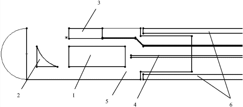
[0028] like figure 1 As shown, the present invention discloses an intravascular photoacoustic ultrasound imaging endoscopic probe excited by high-efficiency collimated light. The endoscopic probe includes: C-Lens1, concave mirror 2, ultrasonic transducer 3, optical fiber 4, and probe housing 5 , and the torsion coil 6. The C-Lens1 is used to collimate the pulsed laser light emitted from the optical fiber 4, and the concave mirror 2 is used to optimize the collimated pulsed laser light and reflect it to the sample to excite ultrasonic signals, which are transduced by the ultrasonic The electrical signal converted by the transducer 3 is transmitted to the acquisition system by the coaxial line, and the ultrasonic transducer 3 can independently complete ultrasonic imaging by sending and receiving.

[0029] The C-Lens1, the concave reflector 2, and the ultrasonic transducer 3 are fixed on the endoscopic probe housing 5; the C-Lens1 is between the optical fiber 4 and the concave r...


PUM

PropertyMeasurementUnit
Diameteraaaaaaaaaa
Radius of curvatureaaaaaaaaaa
Diameteraaaaaaaaaa

Abstract

The invention discloses an intravascular photoacoustic / ultrasonic imaging endoscopy probe of efficient collimation optical excitation. The endoscopy probe comprises a C-Lens, a concave reflecting mirror, an ultrasonic transducer, optical fiber, a probe case and a torque coil; the C-Lens is used for collimating a pulse laser transmitted by the optical fiber. The concave reflecting mirror is used for optimizing the collimated pulse laser and reflecting the collimated pulse laser to a sample to excite an ultrasonic signal. The ultrasonic signal is converted into an electrical signal through the ultrasonic transducer and transmitted to an acquisition system through a coaxial line. The ultrasonic transducer can also independently complete ultrasonic imaging by sending or receiving. According to the intravascular photoacoustic / ultrasonic imaging endoscopy probe of the efficient collimation optical excitation, the C-Lens and the concave reflecting mirror are in use cooperatively, therefore, the pulse laser can be effectively collimated, so that the problem is addressed that lasers focus on one point, the probe deviates from the vascular center, and accordingly image distortion exists and the vascular endoscopy imaging quality is improved.

Description

technical field [0001] The invention relates to the research field of vascular endoscopy, in particular to an intravascular photoacoustic / ultrasonic imaging endoscopic probe excited by highly efficient collimated light. Background technique [0002] In the 21st century, cardiovascular and cerebrovascular diseases have become the number one killer affecting human health, and the rupture of atherosclerotic plaques is the main cause of acute heart disease vascular events. Although there are magnetic resonance angiography, angiography imaging, intravascular Ultrasound imaging, these imaging methods can better present the external shape or internal contour of the blood vessel, but cannot provide local details of the blood vessel wall. Intravascular photoacoustic imaging is a non-invasive and non-ionizing new biomedical imaging method developed in recent years. It has the advantages of both pure photoacoustic imaging and pure ultrasound imaging. The advantages of high penetration...

Claims


Application Information

Patent Timeline
IPC IPC(8): A61B5/00A61B8/12A61B8/08G02B23/24G02B27/30
CPCA61B5/0095A61B8/0891A61B8/12A61B8/4483G02B23/2415G02B23/2438G02B27/30
Inventor 杨思华王磊雷鹏邢达
Owner SOUTH CHINA NORMAL UNIVERSITY
Learn More