Establishment and application of radiotherapy model for in-situ bearing cancer of mouse lung cancer

A mouse, in situ technology is applied in the field of establishing an in situ tumor-bearing model in mice, which can solve the problems of unstable tumor site size, high equipment requirements, and difficult operation, and achieves good application prospects, reliable results, Observe intuitive and convenient effects

Inactive Publication Date: 2018-02-23
SECOND MILITARY MEDICAL UNIV OF THE PEOPLES LIBERATION ARMY
View PDF3 Cites 5 Cited by
  • Summary
  • Abstract
  • Description
  • Claims
  • Application Information

AI Technical Summary

Problems solved by technology

Such methods also have defects such as unstable tumor site and size, difficult operation, and high equipment requirements.
In addition, due to the natur

Method used


Image

  • Establishment and application of radiotherapy model for in-situ bearing cancer of mouse lung cancer
  • Establishment and application of radiotherapy model for in-situ bearing cancer of mouse lung cancer
  • Establishment and application of radiotherapy model for in-situ bearing cancer of mouse lung cancer

Examples


Embodiment 1

[0039] Example 1: Establishment method of an orthotopic tumor-bearing mouse model with normal immune function that can be used to evaluate the effect of radiotherapy

[0040] The research work of this project was approved by the Ethics Committee of the Second Military Medical University.

[0041] The firefly luciferase gene was introduced into the mouse Lewis lung cancer LLC cell line, and the tumor clone cell line stably expressing luciferase was obtained through subcloning screening and expanded. Use DMEM medium to resuspend the recombined LLC and prepare 2×10 6 concentration of the cell suspension for later use. After the C57BL / 6 mice were anesthetized with 10% chloral hydrate, they were placed on the left side of the operating table and fixed. After disinfection, the skin was incised at 10 mm to the right of the xiphoid process of the mouse sternum, and the anterior muscular layer of the right chest wall was peeled off to expose the thoracic wall and intercostal space of...

Embodiment 2

[0053] (1) The orthotopic tumor-bearing model of mice with normal immune function is the same as that in Example 1;

[0054] (2) Feeding of mice: Place the mice in a cage with daily change of litter at 25±1°C to ensure sufficient water and food.

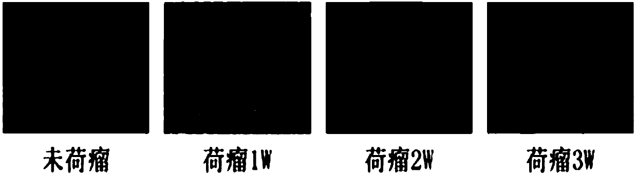
[0055] (3) Three mice were selected respectively before tumor bearing and 1, 2, and 3 weeks after tumor bearing, and after being anesthetized with 10% chloral hydrate, the mice were killed and dissected, and the whole lungs of the mice were taken out. After washing in normal saline, the growth of the tumor was observed and its maximum diameter was measured. Such as figure 2 As shown in the lung tissue observation, it can be seen that the lung tissue of non-tumor-bearing mice is ruddy, smooth and elastic, and no tumor tissue is seen. One week after tumor-bearing mice, obvious tumor tissue can be seen in the lung tissue, with a maximum diameter of about 1 mm, a smooth surface, and a transparent serous interior. Two weeks after bear...

Embodiment 3

[0057] (1) The orthotopic tumor-bearing model of mice with normal immune function is the same as that in Example 1;

[0058] (2) Feeding of mice: Place the mice in a cage with daily change of litter at 25±1°C to ensure sufficient water and food.

[0059] (3) Three mice were selected respectively before tumor bearing and 1, 2, and 3 weeks after tumor bearing, and after being anesthetized with 10% chloral hydrate, the mice were killed and dissected, and the whole lungs of the mice were taken out. After washing in normal saline, the lung lobes with tumor growth were separated, and H&E staining was performed. Such as image 3 As shown, the pathological sections of lung tissue showed that the alveolar wall structure of non-tumor-bearing mice was normal. One week after tumor-bearing mice, pathological sections of the lung tissue showed obvious tumor tissue infiltration, and atypical cells in the tumor tissue grew densely and protruded from the surface of the lung tissue, completel...


PUM


Abstract

The invention relates to the fields of tumor models and establishment and application of tumor models and particularly relates to an establishment method and application a radiotherapy model for mousein-situ bearing cancer. The radiotherapy model can be used for evaluating radiotherapy effects. The establishment method comprises the steps of introducing firefly luciferase genes into a mouse Louislung cancer cell strain (LLC) by virtue of a gene recombination technique by taking a wild mouse as a carrier, carrying out subcloning screening so as to obtain a tumor cloning cell strain capable ofstably expressing luciferase, and inoculating recombinant LLC cells to a lung of the mouse. Compared with a traditional tumor model method, the establishment method has the advantages that the pathological and physiological environments of the lung cancer can be stimulated; the method has very important significances to the diagnosis and treatment of the lung cancer; and the model contains luciferase genes and can be used for imaging in a small animal living body imaging instrument, the development process of the lung cancer can be dynamically monitored in real time, the observation is relatively visible and convenient, living bodies can be observed without injuring the animals, the result is relatively reliable, and the model has very good application prospects.

Description

technical field [0001] The invention relates to the field of tumor model and its establishment and application, specifically, the establishment and application of a mouse orthotopic tumor-bearing model that can be used to evaluate the effect of radiotherapy. Background technique [0002] Lung cancer is the number one killer threatening the health of our people. In the past 50 years, many countries have reported that the incidence of lung cancer has increased significantly. Among male cancer patients, lung cancer has ranked first, and its incidence has also increased rapidly in women, accounting for the second or third of the common malignant tumors in women. The etiology of lung cancer is still not completely clear, but it has seriously affected the life, health and quality of life of patients. Currently, the main treatments for lung cancer include surgery, radiotherapy, chemotherapy and targeted therapy. Among them, radiotherapy is an important local treatment method for ...

Claims


Application Information

Patent Timeline
IPC IPC(8): C12N15/85C12N15/53C12N5/10A01K67/027
CPCA01K67/0271A01K2207/12A01K2227/105A01K2267/0393C12N5/0693C12N9/0069C12N15/8509C12N2510/00C12N2800/107C12Y113/12007
Inventor 高福杨彦勇雷霄蔡建明崔建国李百龙陈媛媛许洋刘哲郭佳铭
Owner SECOND MILITARY MEDICAL UNIV OF THE PEOPLES LIBERATION ARMY
Why Eureka
Try Eureka