Model construction method for cell image classification

A construction method and sub-classification technology, applied in the field of deep learning application research, to achieve high classification accuracy, improve classification accuracy, scale and cell types.

Active Publication Date: 2018-03-23
URIT MEDICAL ELECTRONICS CO LTD +1
View PDF4 Cites 7 Cited by
  • Summary
  • Abstract
  • Description
  • Claims
  • Application Information

AI Technical Summary

Problems solved by technology

[0009] To sum up, although the existing urine formed element cell recognition technology has achieved certain results, most of them have certain limitations, especially

Method used


Image

  • Model construction method for cell image classification
  • Model construction method for cell image classification
  • Model construction method for cell image classification

Examples


Embodiment 1

[0043] Step 1. Image preprocessing

[0044] (1) Divide multiple images in the cell image set A (52000 pieces) into two groups, in which the images with width w and height h satisfying formula (1) form the first group of image sets (28000 pieces), width w and height h The images whose height h satisfies formula (2) form the second set of images (10000):

[0045]

[0046]

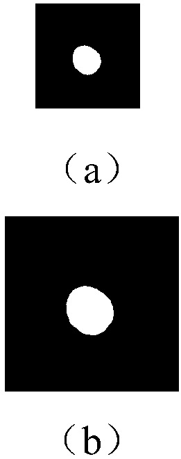
[0047] (2) Classify multiple images in each group of image sets according to the biological characteristics of the cells, and obtain 14 rough classifications in total, carry out subdivisions to each coarse classification, and obtain subdivisions of each coarse classification, a total of 26 subdivisions; The bases for subdivision include cell shape, cell color contrast, and degree of aggregation. For example, for the coarse classification 0-erythrocyte, its subdivision is divided into: normal erythrocytes, nettle-type erythrocytes and wrinkled erythrocytes. The specific classification results are shown ...

Embodiment 2

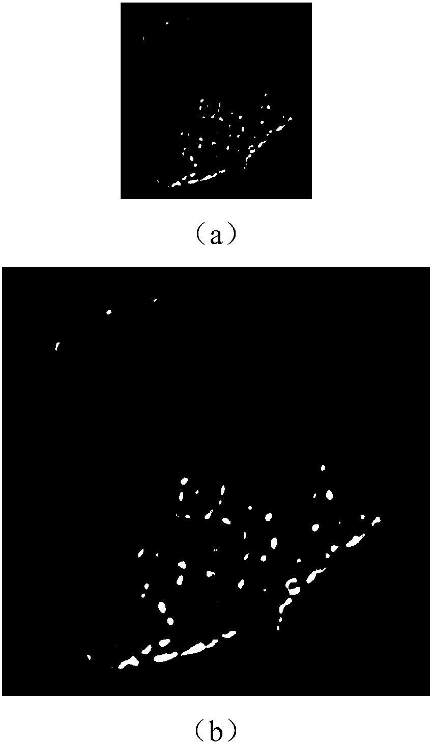
[0060] Utilize the recognition model file of the third group of image collections of embodiment 1 and the recognition model file of the fourth group of image collections to the image to be recognized (such as through step one (1), (3) processing of the present invention image 3 shown) for identification, the specific identification method can use the CLASSIFICATION sample project in the CAFFE framework, the identification result image 3 Cells shown are non-squamous epithelial cells.


PUM


Abstract

The present invention relates to a model construction method for cell image classification. Aiming at the limitations of existing urine sediment recognition technology in real use scenarios, especially the problems that the accuracy is low for multiple classes and the identification speed cannot meet actual production needs, an efficient classification algorithm for urine formed element cells based on multi-model hybrid recognition is proposed. In the present invention, an efficient classification algorithm for urine formed cells based on multi-model hybrid recognition and software implementation include four parts: data preprocessing, obtaining of training model, obtaining of recognition results after threshold control judgment, and image recognition and acceleration .

Description

technical field [0001] The invention belongs to the field of deep learning application research, and relates to a multi-model hybrid recognition algorithm of a deep convolutional neural network and matching recognition software, which can be applied to different types of urine detection instruments and can meet real-time requirements. Background technique [0002] Urine examination has become a widely used clinical examination method because of its simplicity, quickness, and easy access to specimens, and it is one of the current routine clinical examination items in hospitals. [0003] To a certain extent, the type and shape of urine formed cells can reflect the substantial changes in kidney function and the objective expression of some cumulative lesions. For a long time before, people relied on machine shooting and manual selection to process medical images. This method had problems such as low efficiency, high work intensity, and large differences in error with the level ...

Claims


Application Information

Patent Timeline
IPC IPC(8): G06K9/62
CPCG06F18/241G06F18/214
Inventor 王岩宋建锋秦鑫龙蒋均苗启广李东升
Owner URIT MEDICAL ELECTRONICS CO LTD
Why Eureka
Try Eureka