Breeding method for establishing primary biliary cholangitis model mouse through bile duct antigen immunity

A model mouse, antigen immunization technology, applied in the field of medical biology, can solve problems such as serious disease

Inactive Publication Date: 2018-03-27
UNIV OF SCI & TECH OF CHINA
View PDF0 Cites 0 Cited by
  • Summary
  • Abstract
  • Description
  • Claims
  • Application Information

AI Technical Summary

Problems solved by technology

In these mouse models, dnTGFβRII mice and IL-2Rα - / - Mice all have severe systemic autoimmune disease due to genetic modification (J Immunol.2006Aug 1; 177(3):1655-60, Hepatology.2006Nov; 44(5):1240-9), and 2-octyl Alkynoic acid immunized mice have extremely severe peritonitis and late onset (Hepatology.2008 Aug; 48(2):531-40), which largely limit the application of these animal models

Method used


Image

  • Breeding method for establishing primary biliary cholangitis model mouse through bile duct antigen immunity
  • Breeding method for establishing primary biliary cholangitis model mouse through bile duct antigen immunity
  • Breeding method for establishing primary biliary cholangitis model mouse through bile duct antigen immunity

Examples


Embodiment 1

[0052] Example 1. The process of establishing model mice by immunization with bile duct antigens.

[0053] During this process, all mice were wild-type C57BL / 6 background mice purchased from Shanghai Slack Experimental Animal Co., Ltd., and raised in the Specific Pathogen Free (SPF) laboratory animal center of the School of Life Sciences, University of Science and Technology of China. Environment.

[0054] We used collagenase IV (purchased from Sigma-Aldrich, the working concentration of collagenase IV is 0.075% by mass / volume ratio) dissolved in DMEM high-glucose medium (purchased from Thermo Fisher Scientific) to perfuse the mouse portal vein, and the perfusion The temperature was controlled at a constant temperature of 37°C, and after continuous perfusion for 10 minutes (perfusion rate was 5ml / min), the hepatic parenchymal cells attached to the periphery of the bile duct were gently brushed off with a soft brush to obtain a complete bile duct of the mouse. Cut the obtained...

Embodiment 2

[0055] Example 2. Isolation of mononuclear cells in various organs

[0056] Isolation of mononuclear cells from the liver: the liver of the mouse was removed, ground with PBS containing 2% BSA, filtered through a steel mesh, centrifuged at 1500 rpm for 5 min, and the precipitate was taken. The cell pellet was subjected to density gradient centrifugation with 40% percoll (GE Healthcare) for 20 min, and the cell pellet was collected. The erythrocytes in the pellet were lysed for 10 min with the erythrocyte lysate, and the cells were counted under a microscope by a hemocytometer.

[0057] Separation of spleen mononuclear cells: remove the mouse spleen, grind it with two glass slides and PBS containing 2% BSA, filter through nylon mesh, centrifuge at 1500rpm for 5min, and then add red blood cell lysate (purchased from Beyond Biotechnology Co., Ltd. company) lysed the erythrocytes in the cell pellet for 10 min, and then counted the cells under a microscope with a hemocytometer.

...

Embodiment 3

[0060] Example 3. Histopathological examination

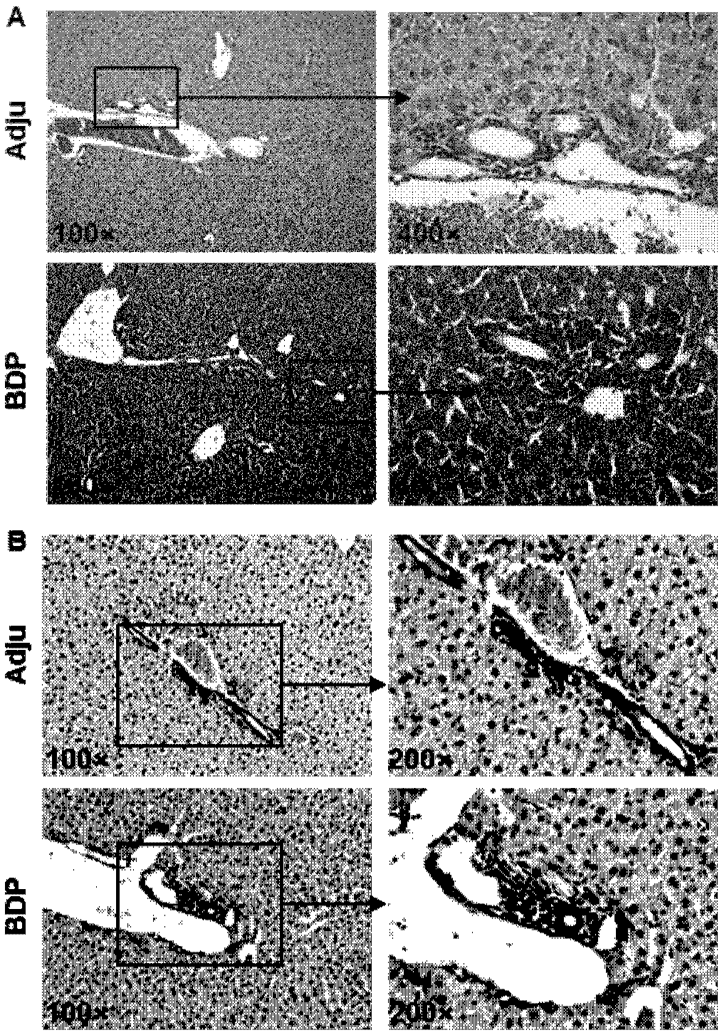
[0061] The mouse liver tissues were fixed in 4% neutral formaldehyde for 1-2 days, dehydrated and embedded in paraffin. Paraffin-embedded liver tissue was sectioned into 4-μm slices, and all slices were deparaffinized and stained with H&E to show infiltration of the portal area in the liver, or deparaffinized and immunohistochemically stained with CK-19 to show inflammation around the bile ducts infiltration.

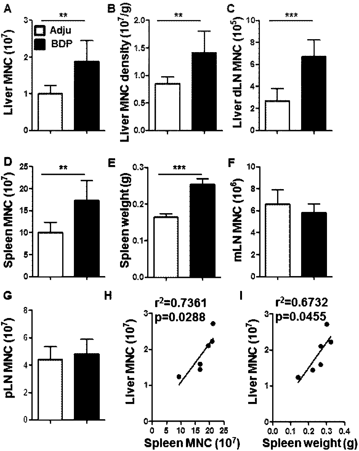
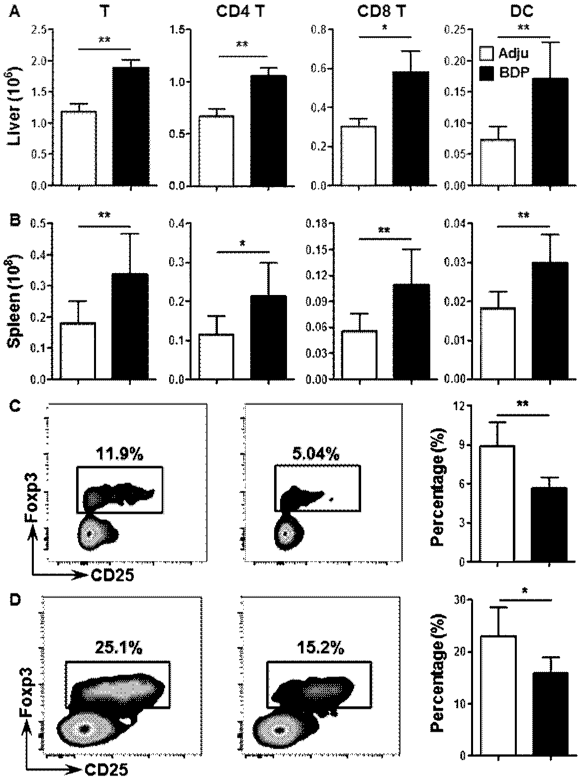
[0062] see results figure 2 HE staining and CK-19 immunohistochemical staining showed that the portal area in the liver of the model mice had obvious inflammatory infiltration, and the specific location of the inflammatory infiltration was around the bile duct. Patients with primary biliary cholangitis usually present with lymphocytic infiltration and bile duct damage in the intrahepatic portal bile duct area, figure 2 The results of the histopathology verified that the performance of the model mice was consistent w...


PUM


Abstract

The invention provides a method for cultivating a mouse model of primary biliary cholangitis, the method comprising: (1) perfusing the mouse liver with collagenase IV for 10 minutes through the hepatic portal vein, removing hepatic parenchymal cells, and obtaining Complete bile ducts; (2) shredding the bile ducts obtained in step (1), further using ultrasonic waves for homogenization, and quantifying the protein concentration of the obtained bile duct protein homogenate; (3) using the obtained bile duct proteins to The source mice were subcutaneously immunized to establish primary biliary cholangitis model mice induced by bile duct antigen immunization. The mouse obtained by the method of the invention can be used as a new animal model for screening drugs for primary biliary cholangitis.

Description

technical field [0001] The invention belongs to the field of medical biotechnology, and in particular relates to a method for cultivating a mouse as a model of primary biliary cholangitis. The mouse obtained by the method of the invention can be used as a new animal model for screening drugs for primary biliary cholangitis. Background technique [0002] Primary biliary cholangitis is a chronic cholestatic disease of the liver. Patients often present with damage to small intrahepatic bile ducts, and high titers of anti-mitochondrial antibodies and inflammatory factors in serum. The disease is female-prone, with a high incidence in middle-aged women. Primary biliary cholangitis can develop from chronic liver inflammatory injury to liver fibrosis, and even further to liver cirrhosis and liver failure (N Engl J Med.2005 Sep 22; 353(12): 1261-73). The disease is difficult to treat effectively, and the only effective drug currently used clinically for the treatment of primary bi...

Claims


Application Information

Patent Timeline
Patent Type & Authority Applications(China)
IPC IPC(8): A01K67/02
CPCA01K67/02
Inventor 廉哲雄马文涛马洪第
Owner UNIV OF SCI & TECH OF CHINA
Why Eureka
Try Eureka