Application of DNA tetrahedron to induction of autophagy
A technology for inducing cells and tetrahedrons, applied to medical preparations containing active ingredients, pharmaceutical formulas, organic active ingredients, etc., can solve the problem of less research on cell physiological activities, and achieve the effect of improving proliferation and migration
- Summary
- Abstract
- Description
- Claims
- Application Information
AI Technical Summary
Problems solved by technology
Method used
Image
Examples
Embodiment 1
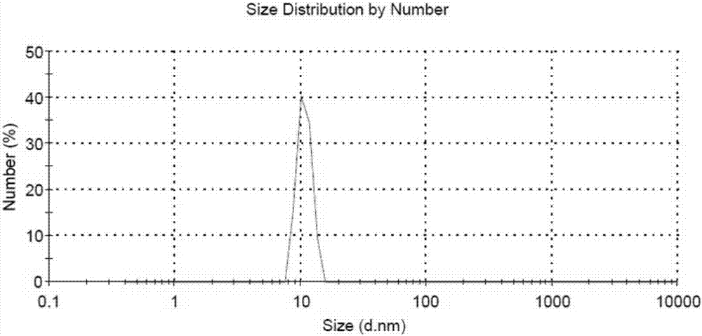
[0048] Synthesis and characterization of embodiment 1 DNA tetrahedron
[0049] 1. Synthesis of DNA tetrahedron
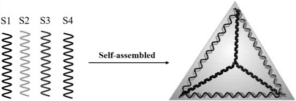
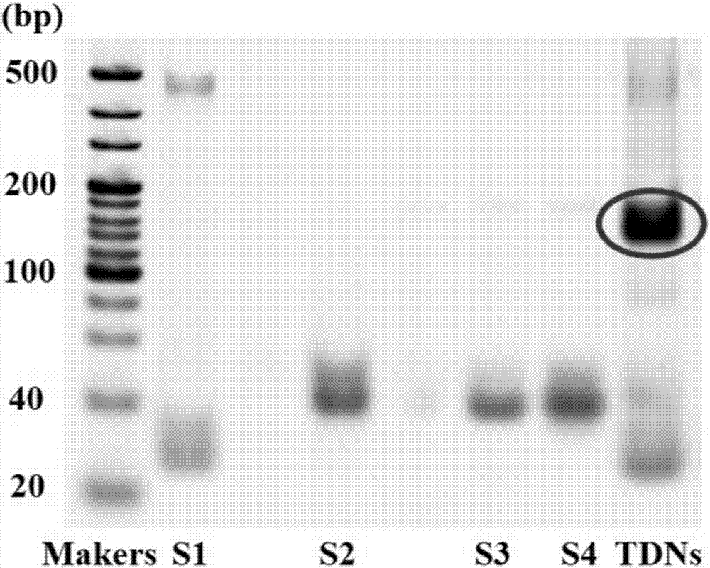
[0050] DNA tetrahedron is composed of uniquely designed four DNA single strands (S1, S2, S3, S4) through a fast, simple and specific PCR program (95°C for 10min, rapid cooling to 4°C for 20min, 4°C for long-term storage) synthesized by self-assembly. The four single strands were added into a 200μl EP tube containing 100ul of TM buffer (10mM Tris-HCl, 50mM MgCl2, pH 8.0) in an equimolar ratio, and the reaction solution was heated to 95°C for 10min, and then rapidly cooled to 4°C to synthesize DNA tetrahedrons body.
[0051] The specific sequences of the four DNA single strands are as follows:
[0052] S1:
[0053] 5'-ATTTATTCACCCGCCATAGTAGACGTATCACCAGGCAGTTGAGACGAACATTCCTAAGTCTGAA-3' (SEQ ID NO: 1);
[0054] S2:
[0055] 5'-ACATGCGAGGGTCCAATACCGACGATTACAGCTTGCTACACGATTCAGACTTAGGAATGTTCG-3' (SEQ ID NO: 2);
[0056] S3:
[0057] 5'-ACTACTATGGCGGGTGATAAAACGTGTAG...
Embodiment 2
[0064] Example 2 Transmission Electron Microscopy Detects the Induction Effect of DNA Tetrahedron on Chondrocyte Autophagy
[0065] 1. In vitro culture of chondrocytes
[0066] The cells used in this experiment were rat chondrocytes, which were obtained from the knee joints of 3-5-day-old SD suckling mice, and the culture medium was high-glucose DMEM containing 10% fetal bovine serum (fetal bovine serum, FBS, Gibco) And 1% double antibody (Penicillin-Streptomycin liquid penicillin and streptomycin mixture). at 37°C, 5% CO 2 Carry out cell culture in an incubator, and when the cells grow to occupy 80-90% of the bottom of the culture dish, they are digested with 0.25% trypsin-EDTA and passaged, and generally transferred to P1-P2 for use.
[0067] 2. Detection of DNA tetrahedron-induced autophagy in chondrocytes
[0068] The ability of DNA tetrahedron to induce autophagy in chondrocytes was detected by transmission electron microscopy. The chondrocytes of the P1 generation we...
Embodiment 3
[0070] Example 3 Confocal microscope detection of LC3 protein expression to observe the induction of DNA tetrahedron on chondrocyte autophagy
[0071] LC3 is a marker protein of autophagy and an important indicator for detecting autophagy. LC3 has two subtypes, namely LC3I and LC3II. The two subtypes switch between each other. When there is no autophagy, the LC3I subtype mainly exists. On the contrary, when autophagy occurs, the LC3II subtype mainly exists. Type exists. Since the LC3Ⅰ subtype protein is water-soluble protein, it will exist in a diffuse state in cells; while the LC3Ⅱ subtype protein is water-insoluble and mainly exists in an aggregated state in water. Therefore, the strength of autophagy in cells can be judged by observing the changes in LC3II content in cells (that is, the distribution of green fluorescence).
[0072] experiment method:
[0073] (1) Digest the chondrocytes of the P1 generation and plant them in a 6-well plate, at 37°C, 5% CO 2 After cultur...
PUM
| Property | Measurement | Unit |
|---|---|---|
| particle diameter | aaaaa | aaaaa |
Abstract
Description
Claims
Application Information
 - R&D
- Intellectual Property
- Life Sciences
- Materials
- Tech Scout
- Unparalleled Data Quality
- Higher Quality Content
- 60% Fewer Hallucinations
Browse by: Latest US Patents, China's latest patents, Technical Efficacy Thesaurus, Application Domain, Technology Topic, Popular Technical Reports.
© 2025 PatSnap. All rights reserved.Legal|Privacy policy|Modern Slavery Act Transparency Statement|Sitemap|About US| Contact US: help@patsnap.com


