
Multiplex PCR (Polymerase Chain Reaction) detection primer group and kit for fast distinguishing porcine deltacoronavirus from porcine kobuvirus

A technology for detecting coronaviruses and primers, which is applied in the field of diagnosis in the field of veterinary biotechnology, and can solve problems such as difficulties in clinical diagnosis

Inactive Publication Date: 2018-05-22
INST OF ANIMAL HUSBANDRY & VETERINARY MEDICINE HENAN ACAD OF AGRI SCI
View PDF1 Cites 3 Cited by
  • Summary
  • Abstract
  • Description
  • Claims
  • Application Information

AI Technical Summary

Problems solved by technology

Because the clinical manifestations and pathological changes of PDCoV and PKoV are very similar, it has caused certain difficulties in clinical diagnosis.

Method used


Image

  • Multiplex PCR (Polymerase Chain Reaction) detection primer group and kit for fast distinguishing porcine deltacoronavirus from porcine kobuvirus
  • Multiplex PCR (Polymerase Chain Reaction) detection primer group and kit for fast distinguishing porcine deltacoronavirus from porcine kobuvirus
  • Multiplex PCR (Polymerase Chain Reaction) detection primer group and kit for fast distinguishing porcine deltacoronavirus from porcine kobuvirus

Examples


Embodiment 1

[0035] 1. Primer design

[0036] According to the PDCoV and PKoV sequences included in GenBank, by comparing the differences between PDCoV and PKoV, design specific primers L1, L2, L3 and L4, as follows:

[0037] L1: 5'-CCTGACACCAACCTGCAGCG-3' (SEQ ID NO.1)

[0038] L2: 5'-GCCCAATGAGTGTACGTCATGCGCTG-3' (SEQ ID NO.2)

[0039] L3: 5'-AGTAGTCCCTACTACTGACGCGT-3' (SEQ ID NO.3)

[0040] L4: 5'-CTACGCTGCTGATTCCTGCT-3' (SEQ ID NO.4)

[0041] Using primers L1, L2, L3 and L4 for PCR amplification, the predicted amplified fragment size of PKoV is 743bp, and the amplified fragment size of PDCoV is 1017bp.

[0042] 2. Extraction of viral RNA

[0043] (1) Take the cell culture fluid of the virus strain, after repeated freezing and thawing 3 times, centrifuge at 5000g for 15min, and take the supernatant for subsequent use;

[0044] (2) Take 0.2 mL of chloroform, add an equal volume of supernatant, shake vigorously for 15 s, leave at room temperature for 3 min, and then centrifuge at 12,00...

Embodiment 2

[0071] The optimization of embodiment 2 primers

[0072] According to the PDCoV and PKoV sequences included in GenBank, by comparing the differences between PDCoV and PKoV, we designed PDCoV-specific primers L5 and L6, as follows:

[0073] L5: 5'-ACATCAGCTGCTACCTCTCCG-3' (SEQ ID NO.5)

[0074] L6: 5'-GGTGGCTCATAGGTCTGGTTAAC-3' (SEQ ID NO. 6)

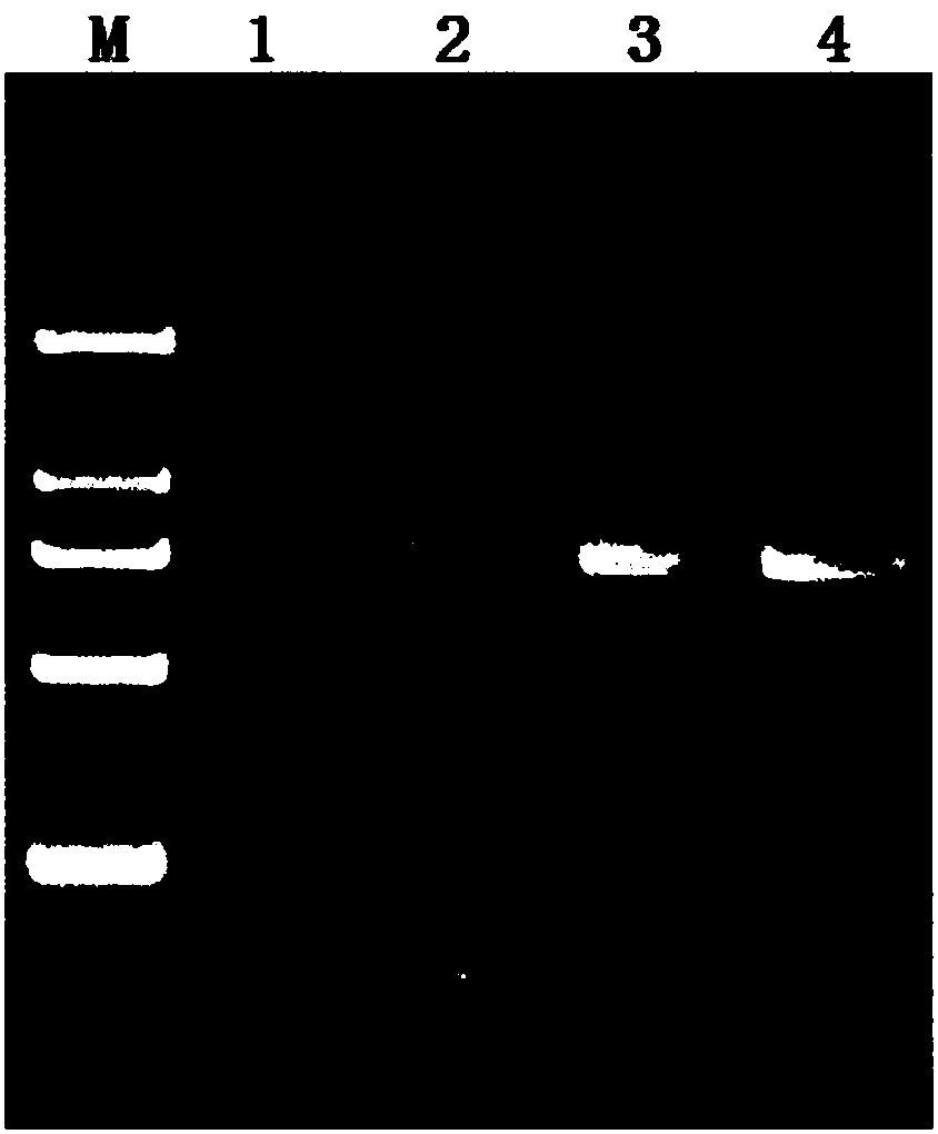
[0075]Using primers L5 and L6 for PCR amplification, the expected amplified fragment size of PDCoV is 516bp. The method of Example 1 was used to amplify, and no target band appeared in the swimming lane. It was found that the annealing temperature of the PCR reaction was 55°C ( Figure 7 ), which is inconsistent with the annealing temperature of PKoV, and cannot be amplified in the same PCR reaction program, so the simultaneous detection of the two viruses cannot be completed.

[0076] Example 3 Amplification Results

[0077] Take PDCoV and PKoV as samples respectively, detect according to the method in embodiment 1, electrophoresis...

Embodiment 4

[0078] Example 4 specificity

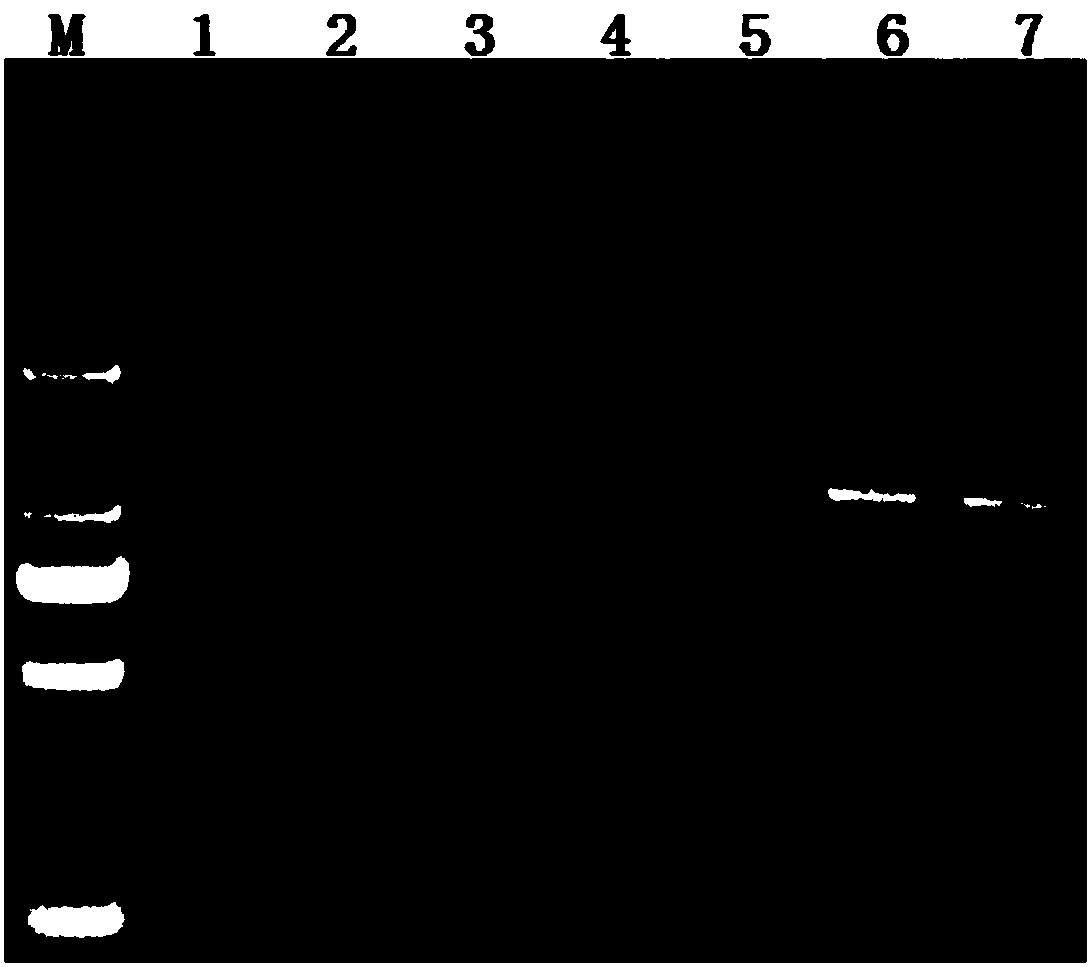
[0079] Porcine circovirus, porcine blue ear disease virus, porcine pseudorabies virus, swine fever virus, porcine parainfluenza virus, porcine parvovirus, and porcine rotavirus were used as control strains, and PDCoV and PKoV were used as experimental strains. The method in example 1 detects ( Figure 9 ), the results showed that only PDCoV and PKoV could obtain 1017bp and 743bp fragments respectively, and no amplification bands were produced by other viruses. Experimental results prove that the primer and detection method of the present invention have high specificity.


PUM


Abstract

The invention discloses a multiplex PCR (Polymerase Chain Reaction) detection primer group and a kit for fast distinguishing porcine deltacoronavirus from porcine kobuvirus. According to PDCoV and PKoV sequences collected in GenBank, differences between PDCoV and PKoV are compared; specific primers L1, L2, L3 and L4 are automatically designed; the primer group can perform specific amplification onPDCoV and PKoV, wherein the primers L1 and L2 ca be used for amplifying PKoV; the primers L3 and L4 can be used for amplifying PDCoV. The inventor successfully invents a special kit on the basis. Virus RNA is firstly extracted; an RT-PCR (Reverse Transcriptase-Polymerase Chain Reaction) method is used for identifying and detecting the PDCoV and PKoV; the size of the PKoV expected amplification fragments is 743bp; the size of the PDCoV expected amplification fragments is 1017bp. Compared with the prior art, the special kit has the advantages of high sensitivity, high specificity and good repeatability, and can be widely applied to clinic PDCoV and PKoV detection.

Description

technical field [0001] The invention relates to a diagnostic technology in the field of veterinary biotechnology, in particular to a multiplex PCR detection primer set and kit for quickly distinguishing porcine delta coronavirus and porcine cristae virus. Background technique [0002] Porcine deltacoronavirus (Porcine deltacoronavirus, PDCoV) is a newly emerged porcine coronavirus, belonging to Nidovirales (Nidovirales), Coronaviridae (Coronaviridae), and Coronavirus (Coronavirus), as a single-stranded positive chain Enveloped RNA virus that was first detected in pigs in Hong Kong in 2012. Subsequently, the virus was also reported to be detected in the United States, Canada, and South Korea. The virus was first reported and detected in mainland China in 2014. Porcine kobuvirus (PKoV) belongs to the family Picornaviridae and belongs to the genus Kobuvirus, and is a single-stranded positive-sense RNA virus. Because the clinical manifestations and pathological changes of PDC...

Claims


Application Information

Patent Timeline
Patent Type & Authority Applications(China)
IPC IPC(8): C12Q1/70C12Q1/686C12N15/11
CPCC12Q1/686C12Q1/701C12Q2537/143Y02A50/30
Inventor 焦文强徐引弟赵珺王克领王治方李宗杰张璐璐孙宗琦王方明郎丽敏周昌龙张立宪游一许峰
Owner INST OF ANIMAL HUSBANDRY & VETERINARY MEDICINE HENAN ACAD OF AGRI SCI
Learn More