Deep learning-based CT image intracranial blood vessel segmentation method and CT image intracranial blood vessel segmentation system

A technology of CT images and intracranial blood vessels, applied in the field of medical image processing, can solve problems such as insufficient accuracy, long algorithm operation time, and low popularity, so as to improve segmentation accuracy, eliminate class imbalance, and accelerate network convergence Effect

Inactive Publication Date: 2018-10-09
上海嘉奥信息科技发展有限公司
View PDF4 Cites 21 Cited by
  • Summary
  • Abstract
  • Description
  • Claims
  • Application Information

AI Technical Summary

Problems solved by technology

[0004] In the field of blood vessel segmentation of CT images, the traditional methods, whether they are based on regions or based on edges, have insufficient accuracy, the scope of the algorithm is very limited, and the robustness to noise is not enough, etc., and some algorithms The operation time is still very long
In particular, for intracranial blood vessel segmentation tasks, these traditional algorithms cannot clearly segment blood vessels in areas where some blood vessels are closely adjacent to the skull.
[0005] As for the emerging deep learning methods, due to the low popularity of the current deep learning methods in the field of medical image analysis, there is no research on the intracranial blood vessel segmentation task of deep learning in CT images.
[0006] Another limitation of deep learning in the field of medical images is that the volume of 3D medical images such as CT is larger than that of ordinary 2D RGB images, and the added dimension makes the convolutional network model require both training and testing. Large video memory to support the calculation of the model, but it is difficult to find a good enough graphics card to support the calculation of the entire CT data

Method used


Image

  • Deep learning-based CT image intracranial blood vessel segmentation method and CT image intracranial blood vessel segmentation system
  • Deep learning-based CT image intracranial blood vessel segmentation method and CT image intracranial blood vessel segmentation system
  • Deep learning-based CT image intracranial blood vessel segmentation method and CT image intracranial blood vessel segmentation system

Examples


Embodiment Construction

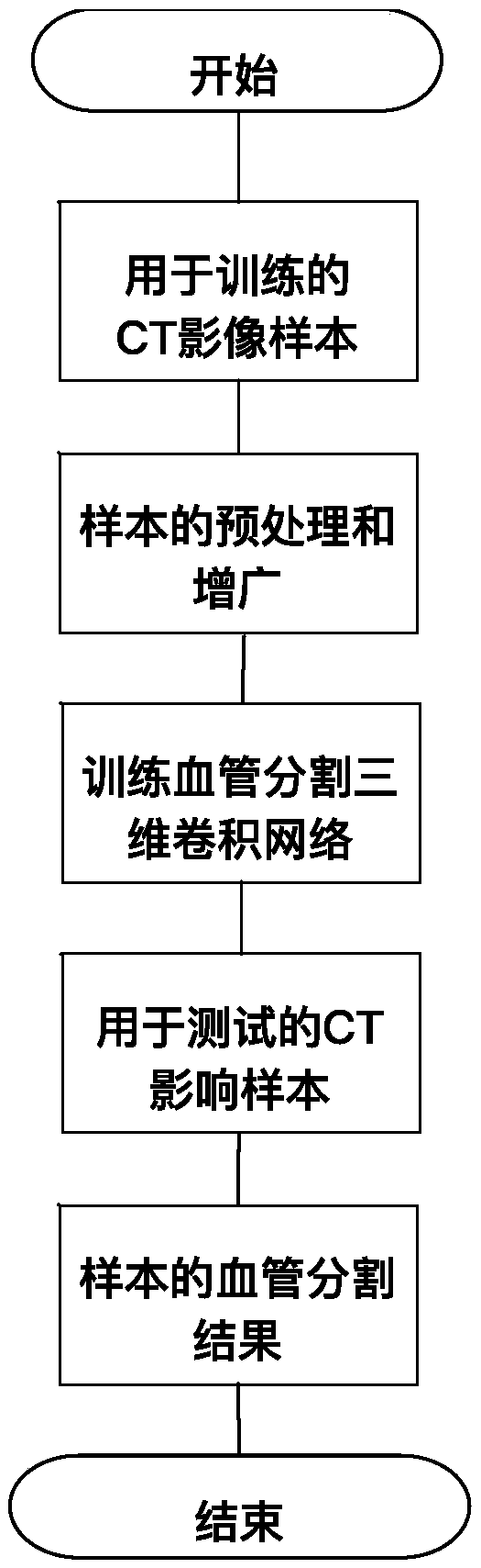
[0038] The present invention will be described in detail below in conjunction with specific embodiments. The following examples will help those skilled in the art to further understand the present invention, but do not limit the present invention in any form. It should be noted that those skilled in the art can make several changes and improvements without departing from the concept of the present invention. These all belong to the protection scope of the present invention.

[0039] Such as figure 1 As shown, a method for intracranial blood vessel segmentation in CT images based on deep learning, including:

[0040] Collection and marking step: collect multiple sets of cranial CTA (CT angiography) data and mark the position of blood vessels, and divide the marked cranial CTA data into training data, verification data and data sets.

[0041] In this example, 70 sets of clinical cranial CTA data were collected, and the blood vessel positions in the data were marked by imaging...


PUM


Abstract

The invention provides a deep learning-based CT image intracranial blood vessel segmentation method and a CT image intracranial blood vessel segmentation system. The method comprises the steps of collecting marks; collecting a plurality of head CTA data sets, marking the positions of blood vessels, and dividing the data sets into a training data set, a verification data set and a test data set; carrying out the preprocessing operation and the augmentation operation: carrying out the preprocessing operation and the augmentation operation on the data sets, wherein the preprocessing operation comprises normalization operation and whitening operation; setting the interlayer spacings of the data sets in three dimensions to be the same through the resampling method, and dividing CT data into small blocks of data according to the size of a preset space window, wherein the augmentation operation comprises the rotating, the amplifying, the reducing and symmetry transformation of data sets; training a three-dimensional convolution neural network: constructing a three-dimensional convolution neural network according to the structure, and training the training data and the verification data according to the parameters. According to the method, the problem that the class of the training process is unbalanced due to the fact that the proportion difference between a background voxel and a vascular voxel is large is eliminated. As a result, the accuracy of the intracranial blood vessel segmentation result is improved.

Description

technical field [0001] The present invention relates to the technical field of medical image processing, in particular to a method and system for segmenting intracranial blood vessels in CT images based on deep learning. Background technique [0002] Vessel segmentation in medical images is the key technology of vascular imaging system, and it is also an important step for three-dimensional visualization of blood vessels, morphological measurement and computer-aided diagnosis. With the development of CT (Computed Tomography, computerized tomography), MRI (Magnetic Resonance Imaging, nuclear magnetic resonance imaging), the development of these advanced imaging technologies and the progress of angiography technology, the vascular images obtained by using them are clearer, and have also prompted many Domestic and foreign scholars have studied in the field of blood vessel segmentation. In recent years, many professional scholars have also proposed many methods, including regio...

Claims


Application Information

Patent Timeline
Patent Type & Authority Applications(China)
IPC IPC(8): G06T7/11G06N3/04
CPCG06T7/11G06T2207/30101G06T2207/20081G06T2207/20084G06T2207/10081G06N3/045
Inventor 吕天予
Owner 上海嘉奥信息科技发展有限公司
Why Eureka
Try Eureka