Probe for in situ nondestructive testing of nucleic acid molecules in extracellular vesicles and preparation method and application thereof

A non-destructive detection and nucleic acid molecular technology, applied in the field of medical detection, can solve the problems of inaccurate detection of EVsmiRNA, lack of protection for the integrity of extracellular vesicles, and leakage of EVs contents, so as to improve detection and screening efficiency and save energy. The effect of shortening the sample size and detection time

Inactive Publication Date: 2018-10-12
CHONGQING UNIV
View PDF5 Cites 5 Cited by
  • Summary
  • Abstract
  • Description
  • Claims
  • Application Information

AI Technical Summary

Problems solved by technology

However, the method of punching holes in the cell membrane will potentially damage the outer membrane of EVs, and there is a risk of leakage of EVs contents, resulting in low final detection results
All of the above methods lack the protection of the integrity of extracellular vesicles, and there is a risk of EVs content leakage, which may lead to low final detection results and cannot accurately detect EVs miRNA

Method used


Image

  • Probe for in situ nondestructive testing of nucleic acid molecules in extracellular vesicles and preparation method and application thereof
  • Probe for in situ nondestructive testing of nucleic acid molecules in extracellular vesicles and preparation method and application thereof
  • Probe for in situ nondestructive testing of nucleic acid molecules in extracellular vesicles and preparation method and application thereof

Examples


Embodiment 1

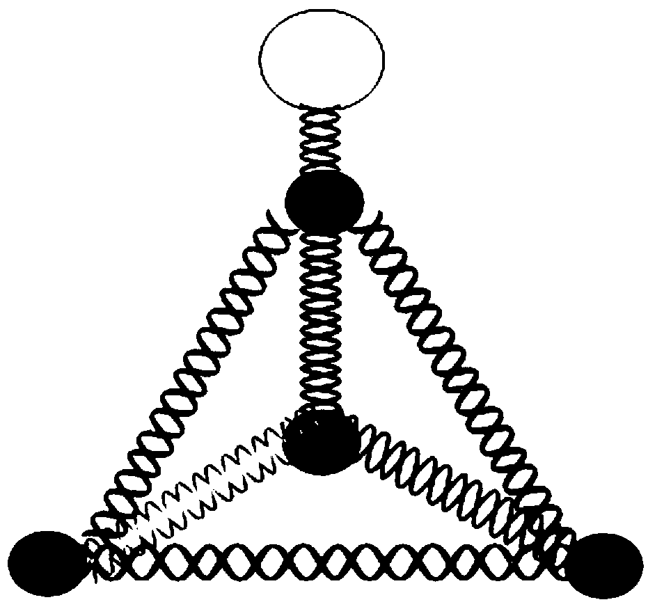
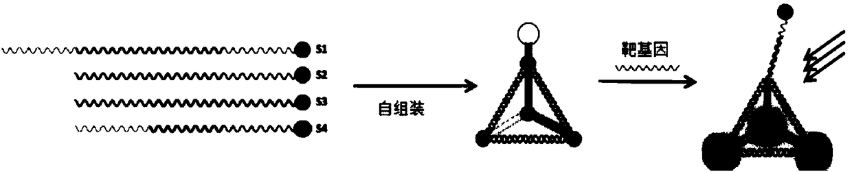
[0032] Example 1. Preparation of tetrahedral probe

[0033] Synthesize 4 single-stranded nucleotide sequences, as follows:

[0034] The first chain: (SEQ ID NO.1);

[0035] The second chain: (SEQ ID NO. 2);

[0036] The third chain: (SEQ ID NO.3);

[0037] Fourth chain: (SEQ ID NO.4);

[0038] The complementary part of each chain in the above sequence uses the same label. The length of the complementary sequence is 13bp, the non-complementary bases are marked in black, the interval between complementary sequences is 2nt, the front of the chain is 1nt, and the bold in the first strand has no complement. Sequence to form a stem-loop probe.

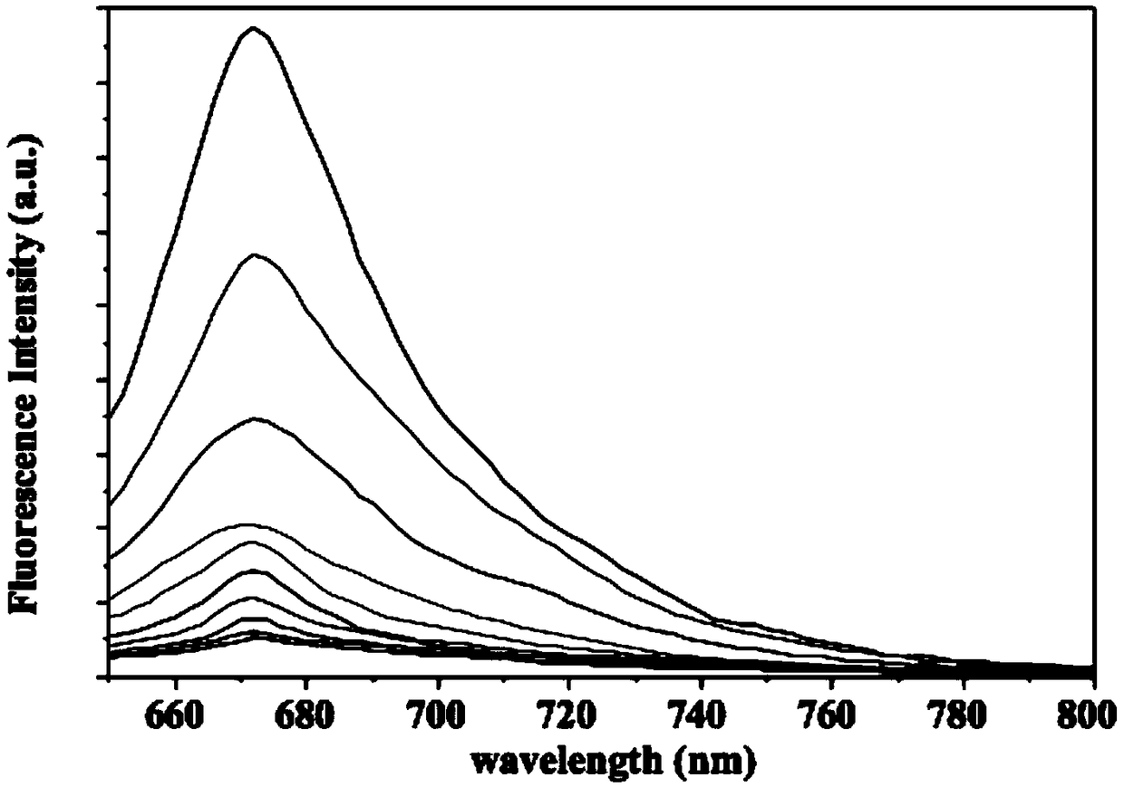
[0039] In order to be able to detect by fluorescence during detection, a fluorescent group was modified at the 5'end of the second, third and fourth chains, and a quenching group was modified at the 5'end of the first chain.

[0040] Dissolve the synthesized single chain in a single-strand self-assembled ion buffer. The preparation method of the sin...

Embodiment 2

[0048] Example 2. Preparation of hexahedral probe

[0049] The nucleotide sequence of synthetically assembled hexahedral probe is as follows:

[0050] First chain 1: (SEQ ID NO.5);

[0051] Second chain 2: (SEQ ID NO.6)

[0052] Third chain 3: (SEQ ID NO.7)

[0053] Fourth chain 4: (SEQ ID NO. 8)

[0054] Fifth chain 5: SEQID NO.9)

[0055] Sixth chain 6: SEQID NO.10)

[0056] The complementary parts of each chain in the six chains use the same label. The complementary length of the hexahedron is 20bp, and the non-complementary bases are black marked with 1nt; the fluorescent group is modified at the 5'of the first, second, third, and fourth chain. At the end, each chain is modified with a fluorescent group; the quenching group is modified at the 5'end of the 5th and 6th chain, and each chain is modified with a quenching group.

[0057] Then the synthesized single strands were self-assembled according to the method in Example 1 to form a hexahedral probe with the structure as Figure...


PUM


Abstract

The invention relates to a polyhedral probe for in situ nondestructive testing of nucleic acid molecules in extracellular vesicles and a preparation method and application thereof. The polyhedral probe comprises a nucleic acid polyhedral skeleton and a probe structure which is used for identifying a target gene and located at a vertex angle of a polyhedron, the probe structure is modified with a quenching group, and the nucleic acid polyhedral skeleton is modified with a luminescent group. The probe can rapidly, sensitively and specifically detect a plurality of nucleic acid molecules in the extracellular vesicles, and can perform design application on the specific target nucleic acid molecules according to the clinical or research detection needs.

Description

Technical field [0001] The invention belongs to the field of medical detection, and relates to a polyhedral probe for in situ non-destructive detection of small nucleic acid molecules of extracellular vesicles, which can quickly, sensitively and specifically detect multiple small nucleic acid molecules of extracellular vesicles with known sequences; The invention also relates to the preparation method and application of the polyhedral probe. Background technique [0002] Extracellular vesicles (EVs) are membranous vesicles secreted by tumor cells and other cells with a size of 50-1000 nm, which are often found in most body fluids such as blood, urine, and cerebrospinal fluid. EVs can carry a variety of intracellular substances. For example, EVs of different sources can contain more than 9,700 proteins, 3,400 mRNAs, and 2,800 microRNAs. They play a role in intercellular signal transmission, tumor occurrence and prognosis monitoring, immune system intervention and other physiologic...

Claims


Application Information

Patent Timeline
IPC IPC(8): C12Q1/6876C12Q1/6806
CPCC12Q1/6806C12Q1/6876C12Q2600/178
Inventor 罗阳刘炼华贾玫陈晓辉张洪邱晓沛卿光超刘春姣王睿璇周旋
Owner CHONGQING UNIV
Why Eureka
Try Eureka