
Ultrasonic scalpel deVice for digestiVe endoscopy

A digestive endoscope and ultrasonic knife technology, applied in the field of medical devices, can solve the problems of inflexibility, too thick diameter, difficult to bend digestive endoscope, etc., and achieve the effect of excellent air barrier property and reduced size

Inactive Publication Date: 2018-10-19
UNIV OF SHANGHAI FOR SCI & TECH
View PDF2 Cites 2 Cited by
  • Summary
  • Abstract
  • Description
  • Claims
  • Application Information

AI Technical Summary

Problems solved by technology

However, the current ultrasonic knife has the disadvantages of being too thick and not bendable, so it is difficult to apply it to the bendable digestive endoscope

Method used


Image

  • Ultrasonic scalpel deVice for digestiVe endoscopy
  • Ultrasonic scalpel deVice for digestiVe endoscopy
  • Ultrasonic scalpel deVice for digestiVe endoscopy

Examples


Embodiment 1

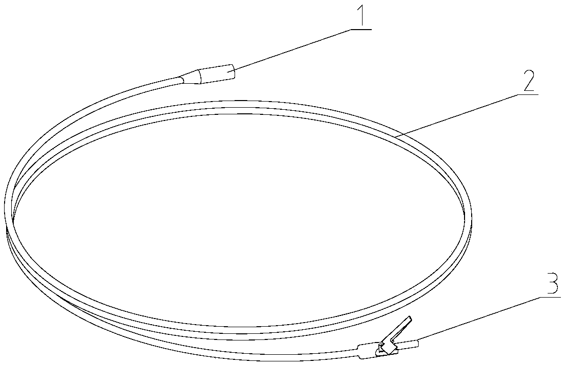
[0026] like figure 1 As shown, an ultrasonic scalpel device for digestive endoscopy includes a main body of an ultrasonic scalpel tube and a control end 1 connected thereto. One end of the shaft 2 is connected to the control end 1, and the other end is connected to the ultrasonic knife head 3; when in use, the main body of the ultrasonic knife intubation enters through the jaw of the digestive endoscope, and enters the operating cavity along the axial direction of the jaw.

[0027] The diameters of the bendable shaft 2 and the ultrasonic cutter head 3 are smaller than the inner diameter of the forceps tube. The flexible shaft 2 is made of soft, oil-resistant, low-temperature shrinkable, and excellent gas barrier materials. In this embodiment, the material of the bendable shaft 2 is soft flame-retardant vinyl fluoride resin. The length of the bendable shaft 2 is 1.5m-2.3m.

[0028] like Figure 8 As shown, the control end 1 is connected with the ultrasonic cutter head 3 thr...

Embodiment 2

[0031] The material of the bendable shaft 2 is polyester, which increases flexibility and controllability, and the length of the bendable shaft 2 is 1.5m-2.3m.

[0032] The ultrasonic knife head 3 is a bipolar electric knife, such as figure 2 shown. All the other are with embodiment 1.

Embodiment 3

[0034] The flexible shaft 2 is a flexible tungsten wire hose, and the inside of the flexible tungsten wire hose is provided with a PET coating.

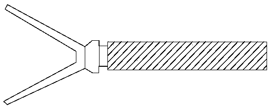
[0035] The ultrasonic cutter head 3 is a separating forceps, such as image 3 shown. All the other are with embodiment 1.

[0036] Wherein the ultrasonic cutter head 3 can also be a variety of multifunctional cutter heads such as grasping forceps, clamp applicator, scissors or coagulator, the diameter of which is smaller than the inner diameter of the tube hole of the forceps, and can complete operations such as cutting, coagulation, grasping, and separation, such as Figure 4-7 shown.


PUM

PropertyMeasurementUnit
Lengthaaaaaaaaaa

Abstract

The inVention relates to an ultrasonic scalpel deVice for a digestiVe endoscopy. The ultrasonic scalpel comprises an ultrasonic scalpel intubation tube body and a control end (1) connected with the ultrasonic scalpel intubation tube body, the ultrasonic scalpel intubation tube body comprises an ultrasonic scalpel head (3) and a flexible shaft rod (2), one end of the flexible shaft rod (2) is connected with a control end (1), and the other end is connected with the ultrasonic scalpel head (3); when the deVice is in use, the ultrasonic scalpel intubation tube body axially enters an operation caVity from a plier channel opening of the digestiVe endoscopy along a plier channel pipe. Compared with the prior art, the ultrasonic scalpel deVice has the adVantages that the diameter is small, the flexibility is high, the deVice can be applied to digestiVe endoscopies and the like, when the deVice is in use, changing a traditional endoscopy is not needed, and it is only required that the ultrasonic scalpel and an endoscope are inserted to conduct obserVation and an ultrasonic scalpel operation synchronously, so that surgical instruments are simplified.

Description

technical field [0001] The invention relates to a medical device, in particular to an ultrasonic scalpel device for digestive endoscope. Background technique [0002] With the rapid development of modern medicine, ultrasonic surgical instruments have been more and more used in the treatment of diseases. It applies ultrasonic energy to surgery. Its outstanding features are fine cutting, safety, tissue selectivity and low temperature hemostasis. The earth has enriched the means of surgery, improved the quality of surgery, and alleviated the pain of patients to a certain extent. The system of a conventional ultrasonic cutting hemostat is mainly composed of a host, a handle, a knife and a foot switch. The host mainly includes an ultrasonic signal generator, a power amplifier and an embedded computer. The low-power ultrasonic electrical signal generated by the ultrasonic signal generator can drive the ultrasonic transducer in the handle to work after being amplified by the powe...

Claims


Application Information

Patent Timeline
IPC IPC(8): A61B17/32
CPCA61B17/320016A61B17/320068A61B2017/320069A61B2017/320074A61B2017/320082
Inventor 侯丽英杨会杰
Owner UNIV OF SHANGHAI FOR SCI & TECH
Learn More