
Lung nodule detection method and device, computer device and storage medium

A detection method, a technology of pulmonary nodules, applied in the computer field, can solve the problems of inability to segment the lung area, slow diagnosis speed, costing doctors, etc., and achieve the effect of improving detection accuracy, fast segmentation speed, and improving the effect

Active Publication Date: 2018-11-06
PING AN TECH (SHENZHEN) CO LTD
View PDF6 Cites 32 Cited by
  • Summary
  • Abstract
  • Description
  • Claims
  • Application Information

AI Technical Summary

Problems solved by technology

[0003] Pulmonary nodules need to be separated from lung CT images before pulmonary nodules are removed. At present, the separation is usually done by relying on the clinical experience of doctors, or by some existing image segmentation algorithms. The diagnosis speed is slow and it costs doctors It takes a long time, and the accuracy rate is low; at the same time, the existing detection method cannot segment the lung area for some special lung CT, which seriously hinders the subsequent detection

Method used


Image

  • Lung nodule detection method and device, computer device and storage medium
  • Lung nodule detection method and device, computer device and storage medium
  • Lung nodule detection method and device, computer device and storage medium

Examples


Embodiment Construction

[0039] In order to make the purpose, technical solution and advantages of the present application clearer, the present application will be further described in detail below in conjunction with the accompanying drawings and embodiments. It should be understood that the specific embodiments described here are only used to explain the present application, and are not intended to limit the present application.

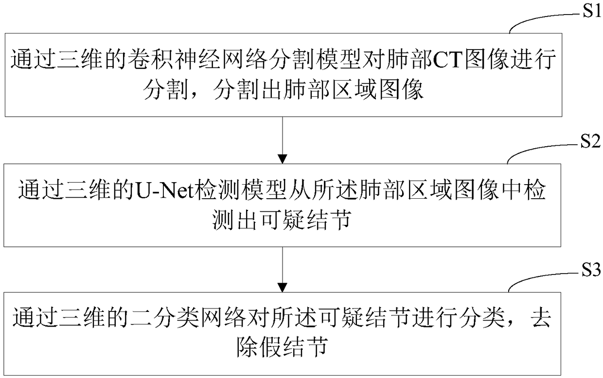
[0040] refer to figure 1 , a method for detecting pulmonary nodules is provided in the embodiment of the present application, comprising the following steps:

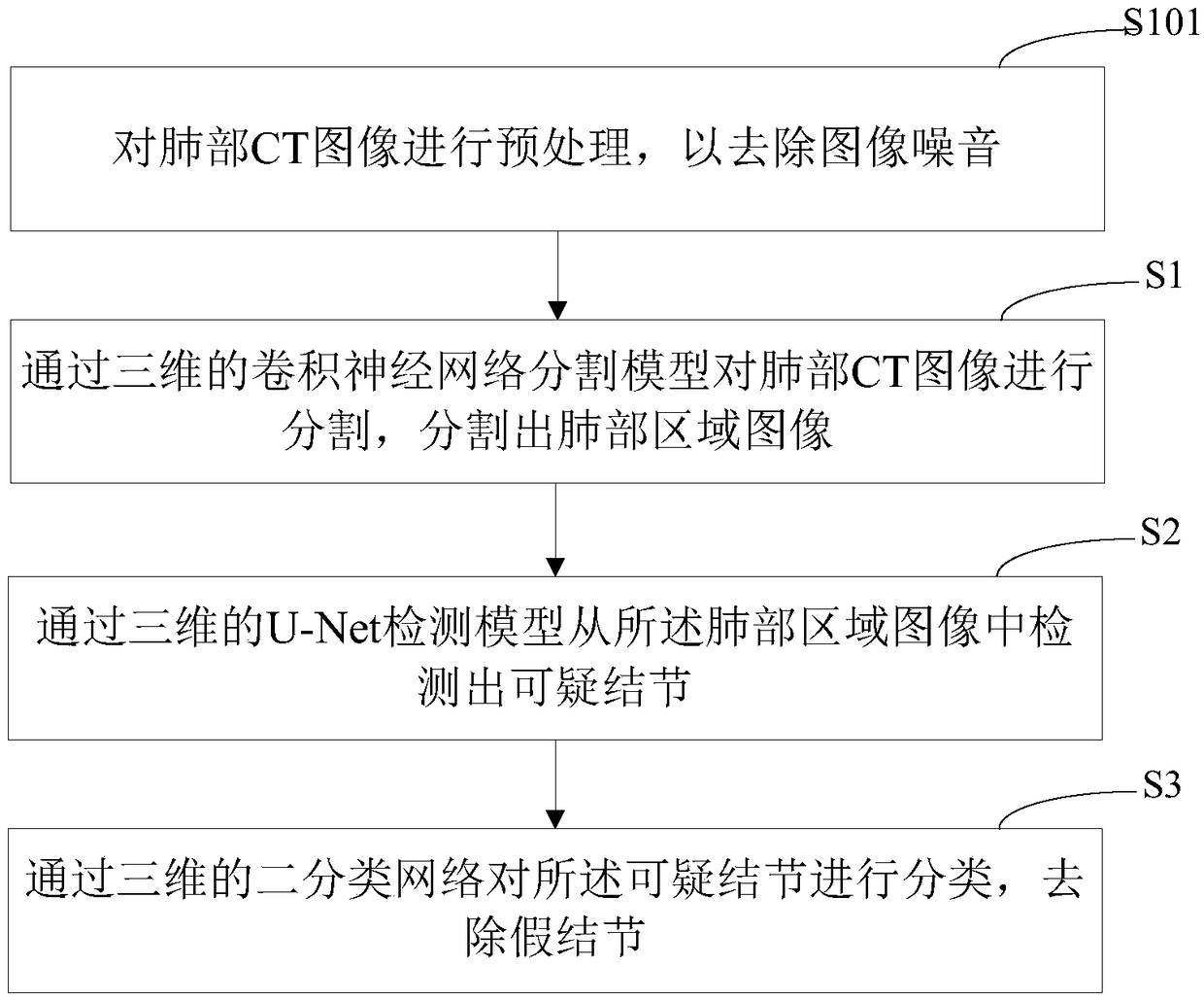
[0041] Step S1, using a three-dimensional convolutional neural network segmentation model to segment the CT image of the lungs, and segment images of the lung region;

[0042] Step S2, using a three-dimensional U-Net detection model to detect suspicious nodules from the image of the lung region;

[0043] In step S3, the suspicious nodules are classified by a three-dimensional binary classification network to remov...


PUM


Abstract

The invention relates to a lung nodule detection method and device, a computer device and a storage medium. The method comprises the following steps segmenting the lung CT image through a three-dimensional convolution neural network segmentation model, and segmenting a lung region image; detecting suspicious nodules from the lung region image through a three-dimensional U-Net detection model; classifying the suspicious nodules through a three-dimensional dichotomous network, and removing the fake nodules. According to the lung nodule detection method and device, the computer device and the storage medium, the lung CT image is segmented through the three-dimensional convolution neural network segmentation model; the lung region image is segmented, the segmentation speed is high, and the speed of subsequent detection of the lung nodules is accelerated; meanwhile, the invention can be applied to the segmentation of the lung region of all lung CT images.

Description

technical field [0001] The present application relates to the field of computer technology, in particular to a detection method, device, computer equipment and storage medium for pulmonary nodules. Background technique [0002] Pulmonary sarcoidosis (sarcoidosis) is a multi-system multi-organ granulomatous disease of unknown etiology, which has attracted widespread attention in China recently. Pulmonary sarcoidosis often invades the lungs, bilateral hilar lymph nodes, eyes, skin and other organs. Pulmonary cell hyperplasia or foreign matter can lead to the development of pulmonary nodules. In the increasingly deteriorating environment, more and more people develop pulmonary nodules in their lungs. Nowadays, pulmonary nodules are a very common symptom, and many young people need to go to the hospital to remove the pulmonary nodules as soon as possible. [0003] Pulmonary nodules need to be separated from lung CT images before pulmonary nodules are removed. At present, the s...

Claims


Application Information

Patent Timeline
IPC IPC(8): G06T7/00G06T7/11G06T5/00G06T17/00G06N3/08G06N3/04
CPCG06N3/084G06T7/0012G06T7/11G06T17/00G06T2207/30064G06N3/045G06T5/70
Inventor 刘新卉刘莉红吴天博马进王健宗肖京
Owner PING AN TECH (SHENZHEN) CO LTD
Learn More